What is a Hypoechoic Mass and What Does It Mean for Your Health?
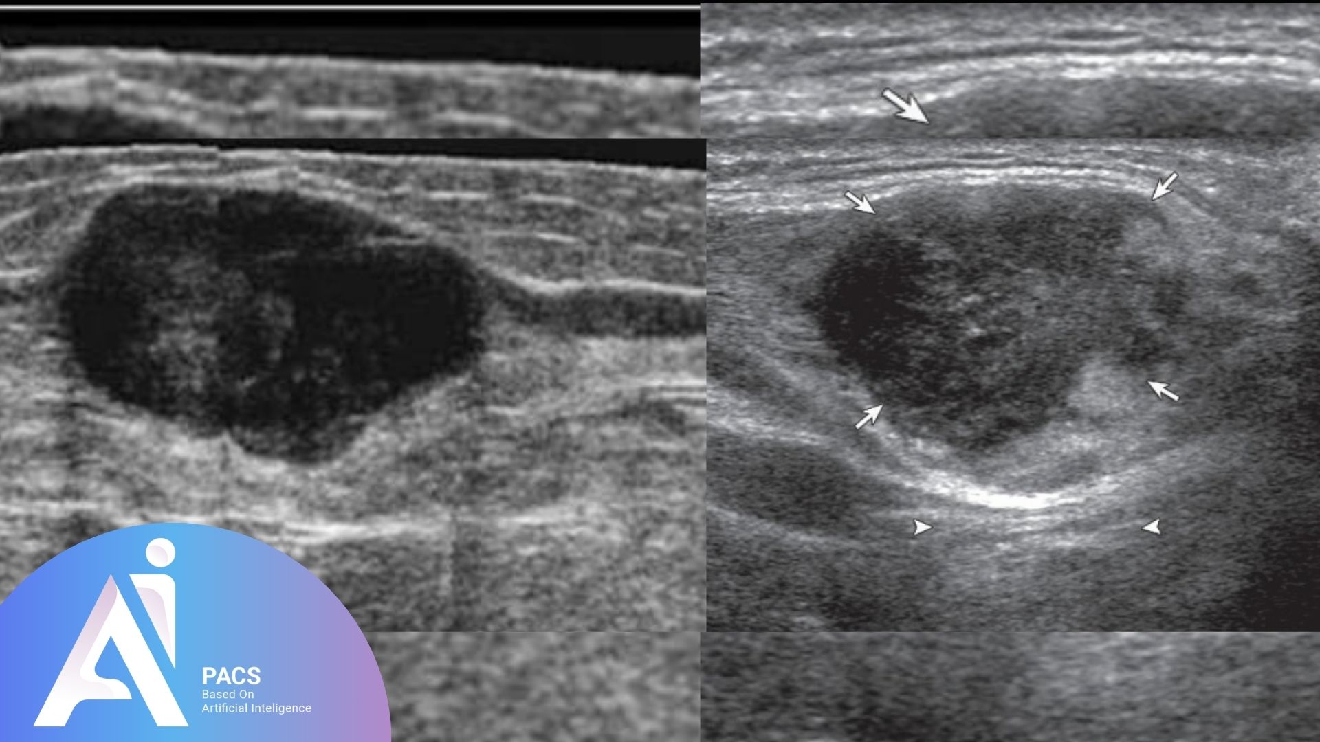
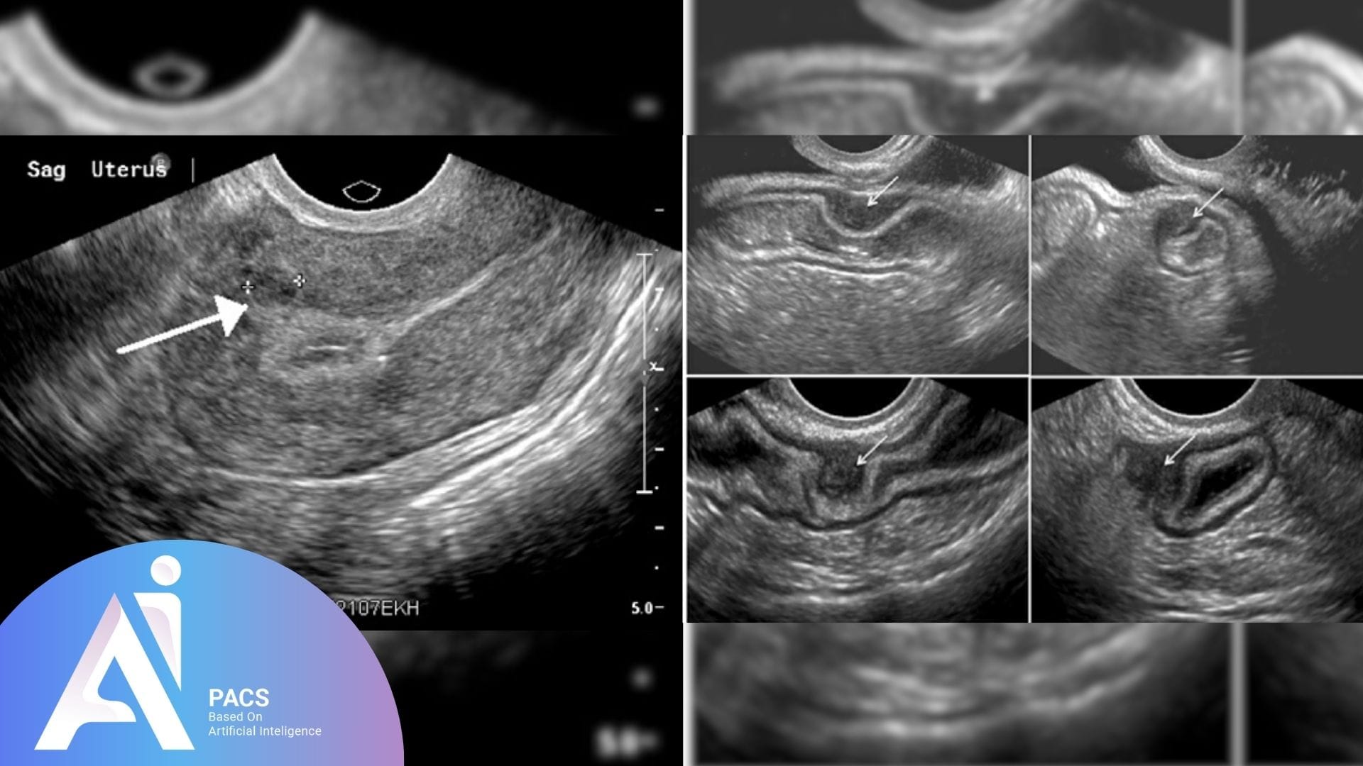
The term “hypoechoic” refers to how this mass interacts with sound waves during an ultrasound exam. It absorbs more sound, reflecting fewer waves to the ultrasound probe. Hypoechoic masses can be solid, semi-solid, or fluid-filled and appear in various organs such as the breast, liver, kidneys, or thyroid. While a hypoechoic mass can be concerning, not all are cancerous, and further testing is often required to determine its nature.
On an ultrasound, a hypoechoic mass is darker than the surrounding tissue due to its reduced reflection of sound waves. In contrast, a hyperechoic mass reflects more sound waves and appears brighter on the screen, while an anechoic mass reflects no sound waves and shows up as a black area, often indicating a fluid-filled cyst. The different appearances of masses help radiologists determine their likely composition and guide further diagnostic steps.
👉 Upload your report now and get an expert review from our online radiology report services.
Why Do Some Tissues Appear Hypoechoic?
Tissues appear hypoechoic when they have a composition that absorbs or transmits sound waves rather than reflecting them. This can happen when the tissue is more solid, dense, or has different acoustic properties than the surrounding tissue. For example, muscle, some tumors, or inflamed tissue can appear hypoechoic. The degree of hypoechoicity can indicate whether the mass is benign or malignant, but further tests like biopsy are usually needed for a definitive diagnosis.
Common Causes of Hypoechoic Masses
Hypoechoic masses can arise in various body parts and may be caused by benign, malignant, or other conditions. The appearance of these masses on ultrasound often necessitates further evaluation to determine their nature and underlying cause.
Benign causes
Many hypoechoic masses are benign, meaning they are non-cancerous and generally pose little health risk. Some common benign causes include:
- Cysts: Although typically fluid-filled and anechoic, some complex cysts may appear hypoechoic due to internal debris or thick walls.
- Fibroadenomas: Common in breast tissue, fibroadenomas are solid, benign tumors that typically appear as hypoechoic masses on ultrasound.
- Lipomas: Fatty tumors, often found in the skin or muscles, can also present as hypoechoic masses and are generally harmless.
Malignant causes
Hypoechoic masses can sometimes indicate malignancy, particularly if they have irregular borders, increased vascularity, or grow over time. Malignant causes may include:
- Breast cancer: Hypoechoic masses in the breast can be a sign of cancer, mainly if they are irregular in shape or show increased blood flow.
- Liver cancer: Hypoechoic masses in the liver may represent primary liver cancer (hepatocellular carcinoma) or metastatic lesions from other cancers.
- Kidney tumors: Hypoechoic masses in the kidneys can be a sign of renal cell carcinoma, a common type of kidney cancer.
Other causes
There are additional causes of hypoechoic masses that do not fall strictly into benign or malignant categories, including:
- Abscesses: Collections of pus caused by infection may appear as hypoechoic masses with irregular borders.
- Inflammatory conditions: Inflammatory masses, such as granulomas, can sometimes appear hypoechoic, particularly in conditions like sarcoidosis or tuberculosis.
- Hemangiomas: These are benign blood vessel tumors that can be hypoechoic on ultrasound, particularly in organs such as the liver.
Understanding the possible causes of a hypoechoic mass is crucial for determining the next steps in diagnosis and treatment, often involving additional imaging or biopsy for confirmation.

What Does Hypoechoic Masses Mean in Different Organs?
The significance of a hypoechoic mass varies depending on the organ in which it is found. Understanding what a hypoechoic mass may indicate in different body parts is essential for proper diagnosis and management.
Breast
A hypoechoic mass in the breast can be a cause for concern, as it may indicate both benign and malignant conditions. Fibroadenomas, which are benign tumors, often appear as well-defined hypoechoic masses. However, irregular, poorly defined hypoechoic masses with increased vascularity could suggest breast cancer. Further diagnostic tools, such as a biopsy or mammogram, are usually required to differentiate between benign and malignant causes.
Liver
A hypoechoic mass in the liver can indicate a range of conditions. Benign lesions, such as hemangiomas or focal nodular hyperplasia, may present as hypoechoic masses, but hepatocellular carcinoma (liver cancer) or metastases from other cancers often appear hypoechoic as well. Liver hypoechoic masses need further evaluation through blood tests, CT scans, or MRIs to determine the exact cause.
Kidneys
Hypoechoic masses in the kidneys can be a sign of both benign and malignant conditions. Simple, standard, and usually harmless cysts may sometimes appear hypoechoic. However, renal cell carcinoma, the most common type of kidney cancer, can also present as a hypoechoic mass. The characteristics of the mass, such as its size and vascularity, help guide whether further testing, like CT or MRI, is needed.
Thyroid
Hypoechoic masses in the thyroid are often associated with thyroid nodules. While many thyroid nodules are benign, specific features, such as irregular margins or microcalcifications, may suggest a higher risk of thyroid cancer. A fine-needle aspiration (FNA) biopsy is often recommended to further evaluate suspicious hypoechoic thyroid nodules.
Uterus/Ovaries
Hypoechoic masses in the uterus or ovaries can indicate various conditions. In the uterus, these masses might represent fibroids (benign muscle tumors). In the ovaries, hypoechoic masses could be benign cysts, such as follicular or corpus luteum cysts, or more concerning conditions like ovarian cancer. These masses’ size, shape, and characteristics, along with patient history, guide further testing and management.
Each organ presents different diagnostic challenges when dealing with hypoechoic masses, making it essential for healthcare providers to perform detailed assessments and use additional diagnostic tools when necessary.
How Is a Hypoechoic Mass Diagnosed?
The diagnosis of a hypoechoic mass typically begins with ultrasound imaging, but further evaluation may involve radiologists’ expertise and additional imaging methods to determine the exact nature of the mass.
The role of ultrasound in diagnosis
Ultrasound is often the first-line imaging tool used to detect hypoechoic masses. It sends high-frequency sound waves into the body, which bounce off tissues and structures, creating an image. Hypoechoic masses absorb more sound than the surrounding tissues, making them appear darker on the ultrasound image. Ultrasound is noninvasive, widely available, and effectively distinguishes between solid and fluid-filled masses. It is invaluable in initially assessing hypoechoic masses in organs like the breast, liver, thyroid, and ovaries.
Role of radiologists in interpreting these masses
Radiologists play a critical role in interpreting ultrasound findings. They analyze the characteristics of the hypoechoic
mass, such as its size, shape, borders, internal structure, and any associated blood flow patterns (through Doppler ultrasound). Based on these features, radiologists assess whether the mass is likely benign or malignant and may recommend further investigation if the mass appears suspicious. Their expert analysis is essential in determining whether the patient needs follow-up imaging, a biopsy, or close monitoring.
Other imaging methods used for further evaluation (CT, MRI)
While ultrasound is often sufficient for an initial evaluation, other imaging methods may be necessary for a more detailed examination. CT (Computed Tomography) scans provide cross-sectional images and are particularly useful for evaluating the internal structure of hypoechoic masses in the liver, kidneys, or other organs. MRI (Magnetic Resonance Imaging) offers high-resolution images and is often used to assess soft tissue masses, providing more detail than ultrasound or CT. CT and MRI can help differentiate between benign and malignant masses, evaluate the extent of any spread (in the case of cancer), and provide a clearer picture before any surgical or non-surgical treatment decisions are made.
When Should You Be Concerned About a Hypoechoic Mass?
While many hypoechoic masses are benign, specific characteristics may raise concerns about the possibility of malignancy. If a mass has irregular borders, rapid growth, or shows increased blood flow on Doppler ultrasound, it could be a sign that further investigation is needed. Additionally, if the mass is found in an area prone to cancer, such as the breast, liver, or thyroid, closer attention and follow-up may be warranted.
The role of biopsy and further tests for confirmation
A biopsy is often the next step when a hypoechoic mass appears suspicious. This procedure involves extracting a small tissue sample from the mass, which is then analyzed under a microscope to determine whether it is benign or malignant. In some cases, a fine-needle aspiration (FNA) is used, particularly in thyroid or breast masses, while larger masses may require a core biopsy. Other tests, such as blood work or tumor markers, may also be performed to support the diagnosis and provide additional information about the nature of the mass.
The importance of follow-up
Regular follow-up is crucial even if a hypoechoic mass is not immediately concerning. Some masses may change, growing larger or developing more suspicious features. Follow-up imaging, such as repeat ultrasound, CT, or MRI, allows healthcare providers to monitor the mass for any size, shape, or appearance changes. In benign cases, consistent monitoring helps avoid unnecessary intervention, while in more concerning cases, early detection of changes ensures timely treatment.
Treatment Options for Hypoechoic Masses
Treatment depends on whether the hypoechoic mass is benign or malignant. Based on diagnostic findings, options range from observation and monitoring to surgery or non-invasive therapies.
Observation and monitoring for benign masses
Observation and regular follow-ups are typically the main approaches for benign masses like cysts or fibroadenomas. No immediate treatment is needed unless there are mass changes, which helps avoid unnecessary surgery.
Surgical or other treatments for cancerous masses
Surgical removal is often the first step when a hypoechoic mass is malignant. For breast cancer, this could mean a lumpectomy or mastectomy. Other organs may require partial removal of the affected area. Surgery may be combined with chemotherapy or radiation therapy to ensure all cancer cells are targeted.
Non-surgical treatments, such as medications or minimally invasive procedures
Non-surgical options, like medications or minimally invasive techniques (e.g., radiofrequency ablation or cryotherapy), can be used for benign or small malignant tumors. These treatments help shrink or destroy masses without the need for major surgery, especially in organs like the liver or kidneys. The treatment approach is tailored to the mass characteristics and patient preferences.
Conclusion
A hypoechoic mass identified on ultrasound can evoke concern, but it’s important to remember that not all such masses are harmful. While some may be benign and require only observation, others may need further investigation through biopsy or advanced imaging techniques. The key to proper management is a thorough evaluation by healthcare professionals, who will guide the next steps based on the characteristics of the masses. Whether the mass is benign or malignant, early detection and appropriate treatment ensure the best possible outcomes for the patient. If you or a loved one encounters the term “hypoechoic mass” in a medical report, consult your doctor for clear guidance and follow-up care.


