A kidney stone CT scan is a specialized imaging test used to detect and diagnose kidney stones, which are hard deposits of minerals and salts in the kidneys. This non-invasive procedure utilizes computed tomography technology to create detailed cross-sectional images of the kidneys and urinary tract, allowing healthcare providers to accurately assess stones’ size, location, and composition. CT scans are particularly valuable because they provide a rapid and precise diagnosis compared to other imaging methods, helping to determine the best treatment options. The procedure typically involves minimal preparation, and while it uses ionizing radiation, the benefits of accurate diagnosis generally outweigh the risks associated with radiation exposure.
kidney stones and the Importance of accurate diagnosis
Accurate diagnosis of kidney stones is crucial for several reasons. First, it helps determine the stones’ size, type (such as calcium oxalate, uric acid, struvite, and cystine stones), and location, which influences treatment options and management strategies. Different kidney stones specialized imaging techniques for accurate diagnosis, as their formation is often linked to specific dietary and metabolic factors.
Second, precise diagnosis can help identify any underlying conditions contributing to stone formation, allowing for targeted interventions to prevent recurrence. Moreover, understanding the severity of the condition enables healthcare providers to promptly address complications, such as obstruction, infection, or severe pain. Finally, accurate diagnosis fosters informed patient decisions, guiding them through treatment options, whether medication, lifestyle changes, or surgical procedures; thereby enhancing overall patient outcomes and quality of life.
What is a CT Scan?
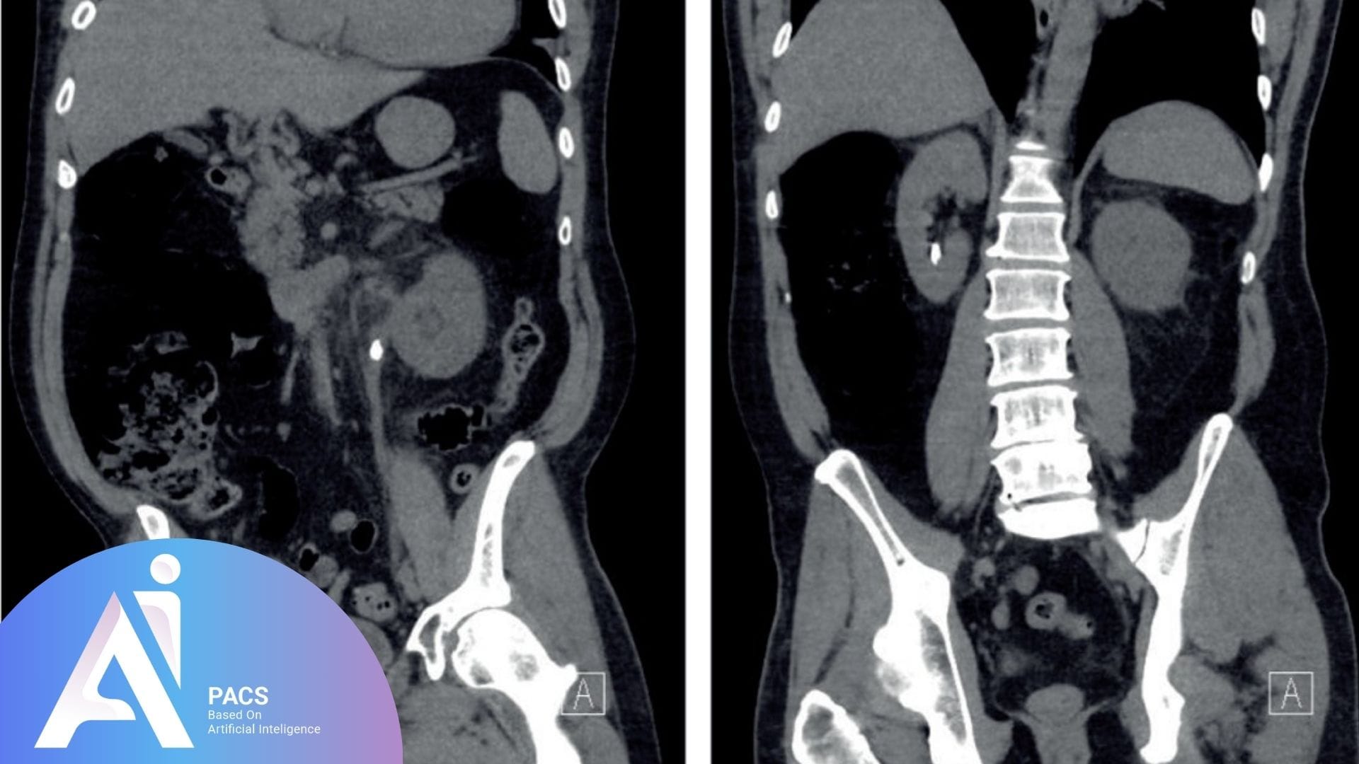
A CT scan, or computed tomography scan, is a medical imaging technique that combines X-ray images taken from different angles and uses computer processing to create cross-sectional images (slices) of the body. This method provides detailed pictures of internal structures, including organs, bones, and tissues, allowing healthcare providers to assess conditions more accurately than standard X-rays. The CT scan can reveal intricate details, making it particularly useful for diagnosing various medical conditions, including tumors, infections, and kidney stones.
How CT scans differ from other imaging techniques (X-ray, ultrasound)
CT scans differ from other imaging techniques like X-rays and ultrasounds in several key ways:
Image Details
X-ray: Provides a two-dimensional image primarily for viewing bone structures and specific organs but lacks detail for soft tissues and complex structures.
Ultrasound utilizes sound waves to produce images, which are excellent for evaluating soft tissue and fluids but may not provide the detailed anatomical views needed to diagnose complex structures like kidney stones.
CT Scan produces highly detailed cross-sectional images, which are superior in visualizing soft tissues and complex structures, including the size and composition of kidney stones.
Speed and Accessibility
X-ray: Quick and widely available, but with limited detail.
Ultrasound: Generally portable and can be done at the bedside but may take longer for comprehensive evaluations.
CT Scan: Usually completed in minutes, providing rapid results, which is crucial in emergencies.
Radiation Exposure
X-ray: Involves some radiation, but usually lower doses than CT scans.
Ultrasound: Does not involve ionizing radiation, making it a safer option for specific populations (e.g., pregnant women).
CT Scans involve higher levels of radiation than standard X-rays, which is why proper preparation is essential, but the detailed information gained often outweighs the risks in appropriate clinical scenarios.
Benefits of using a CT scan for kidney stones
The benefits of using a CT scan for diagnosing kidney stones include:
High Sensitivity and Specificity: CT scans are compassionate, accurately detecting even small rocks that might be missed by X-rays or ultrasounds, thus ensuring a definitive diagnosis.
Detailed Visualization: They provide clear images of the kidneys, ureters, and bladder, allowing for precise assessment of the stone’s size, location, and potential complications, such as obstruction or infection.
Rapid Results: CT scans are typically completed quickly, enabling prompt diagnosis and treatment decisions, which is especially important for patients experiencing severe pain or urinary obstruction.
Comprehensive Evaluation: In addition to diagnosing kidney stones, CT scans can identify other potential issues in the urinary tract, such as tumors, anatomical abnormalities, or associated complications, aiding in overall patient management.
Guiding Treatment Options: The detailed information from a CT scan can help healthcare providers recommend appropriate treatment strategies, whether conservative management, medication, or surgical intervention, tailored to the patient’s specific condition.
Indications for a Kidney Stone CT Scan
Symptoms that may require a CT scan:
Severe Abdominal or Flank Pain: Sudden, intense pain may indicate kidney stones.
Hematuria: Blood in the urine can signal the presence of stones or other urinary issues.
Recurrent Urinary Tract Infections (UTIs): Frequent UTIs warrant investigation for stones, especially with accompanying symptoms, especially with accompanying symptoms, warrant investigation for rocks.
Nausea and Vomiting: These symptoms, alongside abdominal pain, may suggest obstructive stones.
Urinary Obstruction: Suspected obstruction from a stone requires imaging for confirmation.
Suspected Complications: Conditions like hydronephrosis or infection related to stones need evaluation.
History of Kidney Stones: Patients with a previous history and similar symptoms may need a CT scan to diagnose kidney stones.
Assessment Before Treatment: Determining stone size and location helps guide treatment decisions.
Pre-operative Evaluation: Imaging aids surgical planning for stone removal.
Unexplained Abdominal Pain: A CT scan can rule out kidney stones in cases of undiagnosed pain.
The CT Scan Procedure
Preparation before the scan
Preparing for a kidney stone CT scan often involves specific instructions, particularly around diet and hydration, to ensure accurate imaging and patient safety.
Dietary restrictions
Patients may sometimes be asked to avoid eating solid foods for a few hours before the scan, especially if contrast material will be used. This restriction helps reduce the risk of nausea or discomfort and allows for clearer imaging of the abdominal area. Patients should follow their healthcare provider’s guidance on any specific foods or drinks to avoid before the scan.
Hydration requirements
Proper hydration is usually encouraged before a CT scan, as it can help improve image clarity and, if contrast is used, aid in flushing the material from the body afterward. Patients may be asked to drink plenty of water prior to the scan but to avoid other beverages, particularly those that are caffeinated or sugary. Following hydration guidelines can also make the procedure more comfortable and support kidney function.
Step-by-step process of the scan
What to expect during the scan
Arrival and Check-In: Provide any necessary information and complete any required forms.
Preparation: You will be escorted to the scanning room and asked to lie on the CT scanner table.
Positioning: The technician will position you correctly, often on your back, to ensure the best images of your kidneys.
If contrast material is used, it may be injected through an IV or ingested orally, creating a brief warm sensation.
Immobilization: You may be asked to hold your breath briefly during the scan to minimize movement, which can blur images.
Scanning: The table will slowly move through the scanner, and the machine will take a series of X-ray images from various angles.
Post-Scan: Once the scan is complete, you can resume normal activities unless advised otherwise.
Duration of the procedure
The entire CT scan process typically lasts 10 to 30 minutes. The actual scanning portion generally takes about 5 to 15 minutes, depending on the complexity of the exam. After the scan, patients can usually return to their normal activities immediately unless their healthcare provider gives specific instructions.
Use of contrast material (if applicable)
Purpose of Contrast: If a contrast material (dye) is used, it enhances the visibility of blood vessels and tissues, providing more explicit images of the kidneys and urinary tract.
Administration: The contrast can be administered orally or intravenously, depending on the specifics of the scan and what is being evaluated.
Pre-Scan Precautions: Patients should inform their doctor about any allergies, especially to iodine or shellfish, as these may indicate a risk of reaction to the contrast material.
Post-Scan Monitoring: After using contrast, the healthcare team may monitor for any allergic reactions, although severe reactions are rare.
Interpreting the Results
When interpreting the results of a CT scan for kidney stones, several common findings are typically reported:
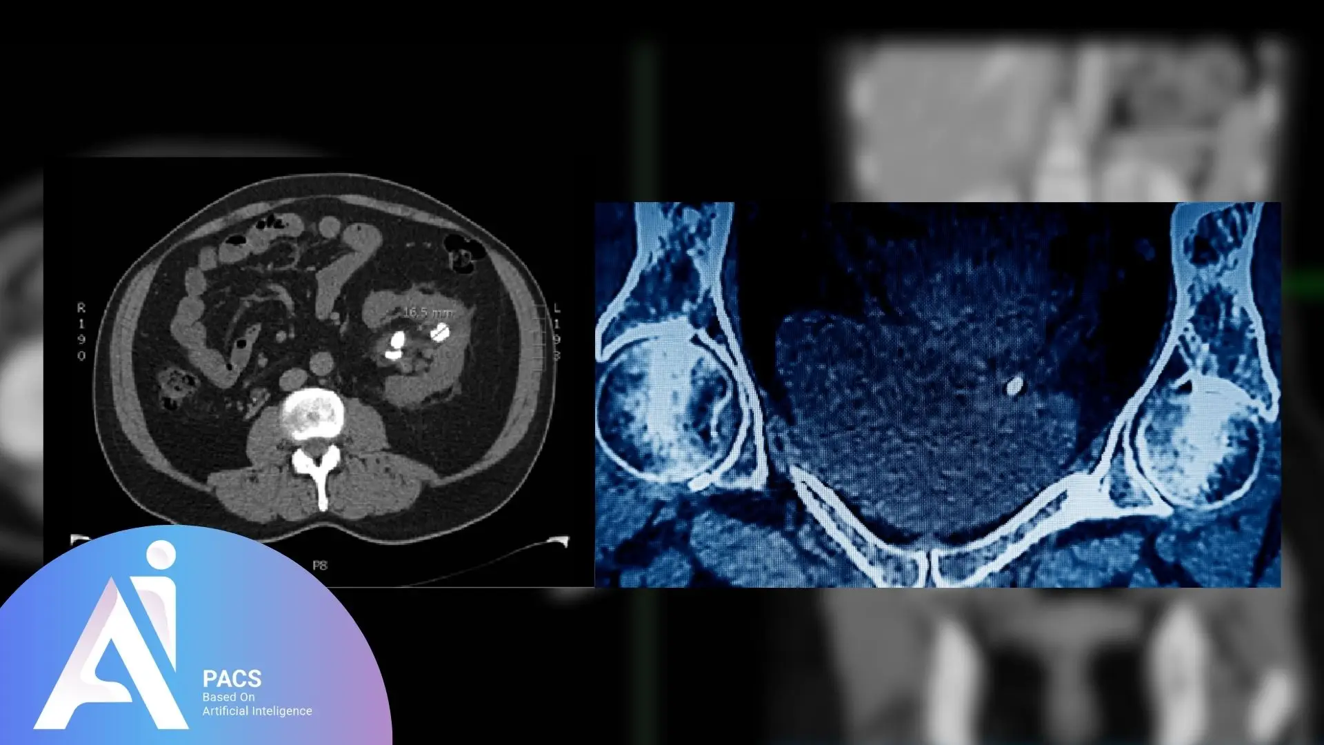
- Presence of Kidney Stones: The scan will confirm the existence of kidney stones, which usually appear as bright white areas due to their density.
- Size of Stones: The report will indicate the size of each stone, measured in millimeters. Larger stones may require different treatment approaches compared to smaller ones.
- Location of Stones: The exact location of the stones will be detailed, specifying whether they are in the kidney, ureter, or bladder. This information is crucial for determining the appropriate treatment.
- Type of Stones: While the CT scan does not directly determine stone composition, specific characteristics may suggest the type of stone (e.g., calcium oxalate stones are denser than uric acid stones).
- Hydronephrosis: This finding indicates kidney swelling due to urine buildup, often caused by a blockage from a stone in the ureter, which may require immediate medical attention.
- Ureteral Dilatation: If the ureter is enlarged, this may indicate an obstruction caused by a stone, further suggesting the need for intervention.
- Associated Complications: The scan may reveal complications such as urinary tract infections (e.g., pyelonephritis) or abnormalities in the urinary tract, which could influence treatment decisions.
Understanding these findings helps guide the management and treatment options for kidney stones, ensuring effective patient care. While these findings provide valuable diagnostic information, interpreting CT scan results requires specialized medical expertise. If you’re uncertain about your kidney stone CT findings or need clarification on treatment options, professional radiology consultation can provide peace of mind and ensure you understand your diagnosis completely.
Want to Understand Your CT Scan Report Better?
After undergoing a CT scan for kidney stones, it’s important not just to wait for a diagnosis—but to understand what your report actually says. CT scan reports can be filled with medical terms that may seem overwhelming at first glance. Knowing how to read and interpret the structure of a typical CT report helps you engage more confidently with your doctor and make informed decisions.
Explore our professional CT scan report interpretation service for personalized analysis.
Post-Scan Follow-Up
Post-scan follow-up is a crucial step after undergoing a CT scan for kidney stones, as it allows for the review and interpretation of the scan results. Following the scan, patients can typically resume normal activities unless directed otherwise. If contrast material is used, healthcare providers may monitor patients briefly to ensure no adverse reactions. The images are analyzed by a radiologist, who prepares a report detailing any findings, which will be discussed during a follow-up appointment with the patient’s healthcare provider.
During this follow-up visit, patients can discuss the results and their implications for treatment. The healthcare provider will explain what the findings mean about the patient’s symptoms and medical history and outline the next steps in management. Depending on the scan results, treatment options may range from conservative measures, such as increased hydration and pain management, to more invasive procedures for larger stones. This follow-up provides clarity and direction for treatment and addresses any questions or concerns patients may have regarding their condition and future management.
If you need help understanding your kidney stone CT results, our expert radiologists provide detailed interpretations to guide your treatment decisions.
Conclusion
In conclusion, a CT scan for kidney stones is an essential diagnostic tool that provides invaluable insights into the presence, size, and location of stones within the urinary tract. Understanding the procedure, the potential risks and the implications of the results equips patients with the knowledge they need to engage actively in their healthcare. Effective communication with healthcare providers is crucial for determining the most appropriate treatment options and managing kidney stone health, from initial diagnosis to post-scan follow-up. By being informed about the entire process from preparation to interpretation of results. Patients can take proactive steps toward their recovery and adopt preventative measures to reduce the risk of future stones, ultimately leading to better health outcomes and improved quality of life.
