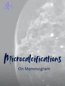

What Are Calcifications?
Calcifications are tiny deposits of calcium that form in the breast tissue. They appear as white spots or flecks on a mammogram and are a common finding, especially in women over 50. Calcifications are calcium deposits, not a diagnosis of cancer. Their shape, distribution, and changes compared with previous mammograms help the radiologist decide whether more evaluation is needed. There are two main types:
Macrocalcifications: Large, coarse deposits usually benign and unrelated to cancer.
Microcalcifications: Tiny deposits that can occur in benign conditions or alongside breast cancer. Small size or clustering alone does not establish a diagnosis. For more detail, read our guide to microcalcifications on mammograms.
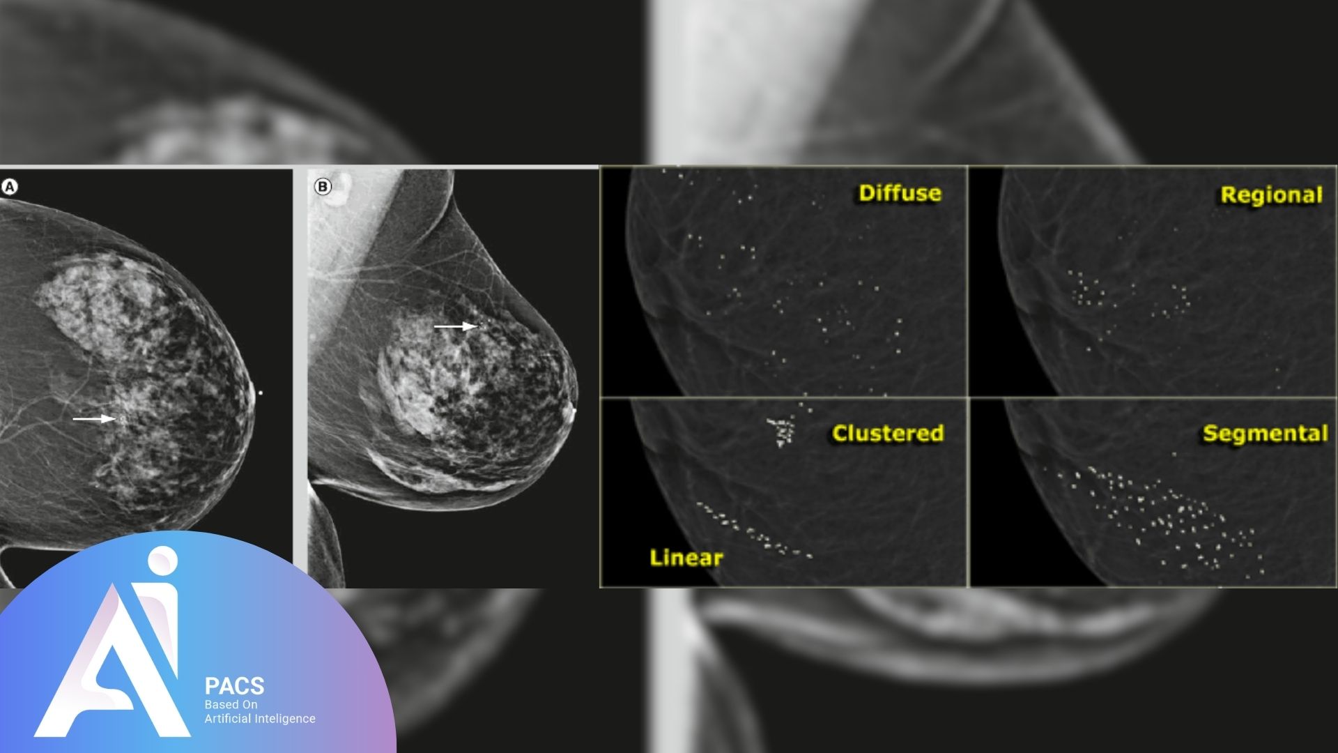
Differentiating between microcalcifications and macrocalcifications:
The terms describe the size of the deposits. Their appearance and the overall assessment determine the next step.
Macrocalcifications
Size: Larger, coarse calcium deposits.
Appearance: Scattered or spread out in the breast tissue, typically easy to identify.
Common Causes: Normal aging, previous breast injury, inflammation, or benign conditions like cysts.
Significance: Usually benign. When the radiologist identifies a typically benign pattern, no additional work-up of those calcifications is generally needed. Continue the screening plan appropriate for you.
Microcalcifications
Size: Smaller, fine calcium deposits.
Appearance: Can appear as clusters or irregular patterns.
Common Causes: These may be associated with benign conditions or indicate more concerning changes, such as Ductal Carcinoma In Situ (DCIS) or early-stage breast cancer.
Significance: This requires closer evaluation, and if the pattern is suspicious, further diagnostic steps, like a biopsy, may be recommended to rule out cancer.
Causes of Calcifications
Calcifications can have benign or malignant causes.
Benign causes
Aging: Natural changes in breast tissue over time.
Previous breast injuries: Trauma or surgery can lead to calcium deposits as the tissue heals.
Infections: Conditions like mastitis can result in calcifications.
Benign breast conditions like fibroadenoma or cysts: Non-cancerous growths like fibroadenomas or cysts often lead to calcifications.
Radiation Therapy: Previous radiation treatment can leave calcium deposits in breast tissue.
Potentially suspicious causes
Ductal carcinoma in situ (DCIS): A non-invasive form of breast cancer often associated with clustered microcalcifications.
Invasive Breast Cancer: Some patterns of calcifications may suggest early signs of invasive cancer.
Atypical Hyperplasia: Abnormal cell growth in breast ducts or lobules may lead to suspicious calcifications.
How Are Calcifications Detected?
Calcifications are usually identified on screening or diagnostic mammograms. Diagnostic imaging investigates a specific finding; its timing follows the recommendation in your report.
Role of mammograms in detecting calcifications
Mammograms are specialized X-ray images of the breast that play a crucial role in detecting calcifications. During the procedure, the breast is compressed, and X-rays are taken, highlighting areas of dense tissue and revealing any calcium deposits. Calcifications appear as small white spots or dots on the mammogram, and their size, shape, and distribution can provide important information about their nature.
Importance of radiologist expertise in interpreting calcification patterns
Interpreting calcifications requires specialized training and experience. Radiologists analyze the mammogram for specific patterns of calcifications to determine their significance:
- Benign vs. Suspicious Patterns: Radiologists look for characteristics such as clustering, shape, and size to differentiate between benign macrocalcifications and potentially concerning microcalcifications.
- Contextual Analysis: To provide a comprehensive assessment, the doctor considers the patient’s medical history, risk factors, and previous imaging results.
- Follow-up Recommendations: Based on their interpretation, radiologists may recommend additional imaging, such as diagnostic mammograms or ultrasounds, or further evaluation through biopsy if suspicious calcifications are identified.
This expertise is essential for accurate diagnosis and appropriate management of breast health.
Calcification Patterns and Their Significance
Benign calcification patterns
Scattered Macrocalcifications: Large, coarse calcium deposits spread randomly throughout the breast tissue. These are common in aging and benign conditions and require no further action.
Popcorn Calcifications: Large, irregularly shaped calcifications that develop in benign growths like fibroadenomas as they age. They have a distinctive “popcorn” appearance and are unrelated to cancer.
Vascular Calcifications: Calcifications that form along the blood vessels within the breast. These are benign and often seen in older women, especially those with diabetes or heart disease.
Ring-Like (Eggshell) Calcifications: Thin, circular deposits surrounding cysts or benign masses, usually indicating non-cancerous conditions.
Suspicious calcification patterns
Clustered Microcalcifications: Small calcium deposits that appear closely grouped. A cluster can be benign or suspicious. The radiologist evaluates the shape of the deposits, their distribution, and changes over time before recommending follow-up or biopsy.
Linear or Branching Calcifications: Microcalcifications arranged in a line or branching pattern may suggest the presence of abnormal cell growth within the milk ducts, potentially indicating a malignancy.
Heterogeneous or Irregularly Shaped Calcifications: These calcifications vary in size and shape and may be associated with more concerning underlying processes, warranting further investigation.
Suspicious patterns often require additional imaging, closer monitoring, or a biopsy to determine whether they are associated with cancerous changes.
What Happens if Calcifications Are Detected?
When calcifications are found during a routine mammogram, the next steps depend on their characteristics. No immediate action may be needed if they appear benign, but suspicious calcifications often require further evaluation. Below are more detailed explanations of the diagnostic steps that may follow the detection of calcifications.
Additional mammogram views (magnified images for closer evaluation)
Purpose: To get a more detailed view of the calcifications. Radiologists may request additional views if initial mammogram images show microcalcifications that appear clustered or have irregular shapes.
Procedure:
- A diagnostic mammogram is performed, which includes taking magnified images of the specific area where the calcifications are seen.
- These zoomed-in images allow radiologists to closely examine the calcifications’ size, shape, and distribution.
Outcome: This helps determine whether the calcifications are benign or suspicious, which guides the need for further testing or a biopsy.
Where ultrasound and MRI fit
Ultrasound may help assess an associated lump or other finding, but some calcifications seen on mammography are not visible on ultrasound. MRI may be used for a separate clinical indication. Neither a normal ultrasound nor a normal MRI should be used on its own to dismiss a mammographic biopsy recommendation. Ask your breast imaging team which test addresses the finding in your report.
Biopsy (if calcifications appear suspicious for cancer)
Purpose: If the completed mammographic assessment is suspicious, biopsy may be recommended to identify the underlying tissue change. Clustering alone does not determine the need for biopsy.
Types of Biopsies:
- Stereotactic Needle Biopsy:
- This is a common method used to evaluate suspicious calcifications. The biopsy is guided by mammography, and a needle removes small tissue samples from the area where the calcifications are located.
- Procedure: Depending on the equipment, the patient may lie face down or sit upright, and the breast is positioned and compressed. The radiologist uses X-rays to precisely guide the needle to the area of concern and extracts tissue samples for analysis.
- Surgical Biopsy:
- In some cases, a surgical biopsy (excisional biopsy) may be needed if the area is difficult to access with a needle. This procedure removes the entire area of suspicious calcifications for a more thorough examination.
Outcome: A pathologist examines the tissue under a microscope to determine whether cancerous or pre-cancerous cells are present. If the results are benign, regular follow-ups may be suggested. If cancer is detected, appropriate treatment options are discussed.
Each step aims to provide a clearer picture of the nature of the calcifications, ensuring that any necessary follow-up or treatment is timely and appropriate.
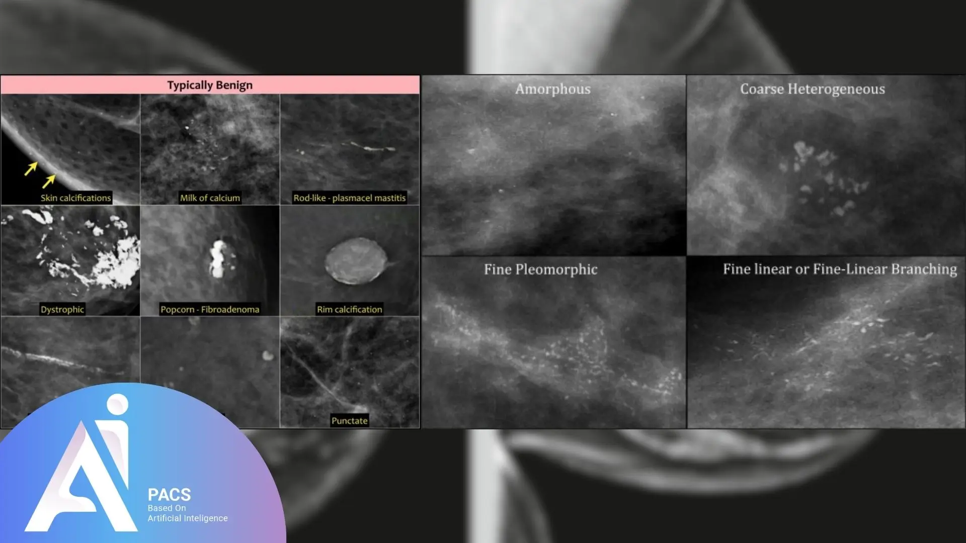
Benign vs. Suspicious Calcifications
A description of size alone does not determine whether a finding is benign. Some tiny calcifications have a typically benign appearance, while others need additional assessment. The combination of shape, distribution, previous images, and the final BI-RADS recommendation matters.
Do not use a picture or a list of patterns to diagnose your own mammogram. If the report recommends biopsy, an image review can help explain that recommendation but does not replace tissue sampling.
Screening and Follow-Up After Calcifications
Routine screening and follow-up for a finding are different. If your report recommends additional views, a short-interval mammogram, or a biopsy, follow that plan rather than waiting for your next screening visit.
Your BI-RADS assessment and recommended next steps help explain the plan. BI-RADS 0 means the assessment is incomplete; BI-RADS 2 describes a benign finding; BI-RADS 3 usually calls for short-interval follow-up. BI-RADS 4 or 5 generally leads to a biopsy recommendation.
Routine screening depends on your age, risk, and local guidelines. For example, the US Preventive Services Task Force recommends screening every two years for women ages 40 to 74 in the population covered by its guidance. Your clinician may recommend a different plan based on your history.
Questions to Ask About Your Calcification Report
- Are the calcifications described as benign, probably benign, or suspicious?
- Which breast and area are involved, and have they changed from previous images?
- Are magnification views needed before the assessment is complete?
- What follow-up or biopsy is recommended, and when should it happen?
If you already have a report and want an independent reread, see how a mammogram second opinion works. For an image review, provide the complete original DICOM study, the existing report, and previous mammograms when available. A report PDF or screenshot alone is not enough for a diagnostic image interpretation. Keep scheduled imaging, biopsy, and treatment appointments while a review is being arranged.
Conclusion
Understanding calcifications in mammography is critical to making informed decisions about breast health. While most calcifications are associated with benign changes, some patterns may indicate the need for further evaluation to rule out or confirm more severe conditions like early-stage breast cancer. Regular mammograms, combined with the expertise of your healthcare provider, help ensure that any concerning findings are identified and addressed promptly. Staying informed and proactive in following up on your mammogram results is essential to maintaining breast health and ensuring early detection when necessary.
Ready to request an independent review of an existing mammogram? Start your mammogram second-opinion request. If you have images but no report yet, read about online radiology reporting to choose the appropriate service.
References
- Canadian Cancer Society: Breast calcifications
- ACR/RSNA RadiologyInfo: Stereotactic breast biopsy
- American Cancer Society: Understanding your mammogram report
- USPSTF: Breast cancer screening
Prefer a quick visual version? See breast calcifications explained (visual story).