





Understanding MRI Terminology
Magnetic Resonance Imaging (MRI) is a powerful tool that provides detailed images of the body’s internal structures. While reading an MRI report can be complex for those without a medical background, understanding its basics can help patients have informed discussions with their healthcare providers. This guide breaks down the essential elements of an MRI report, making it easier to understand what is being described.
If you have MRI images or an MRI report, please don’t hesitate to contact us for a consultation.
Reading an MRI report often involves understanding a series of technical terms. Here’s a list of some common ones:
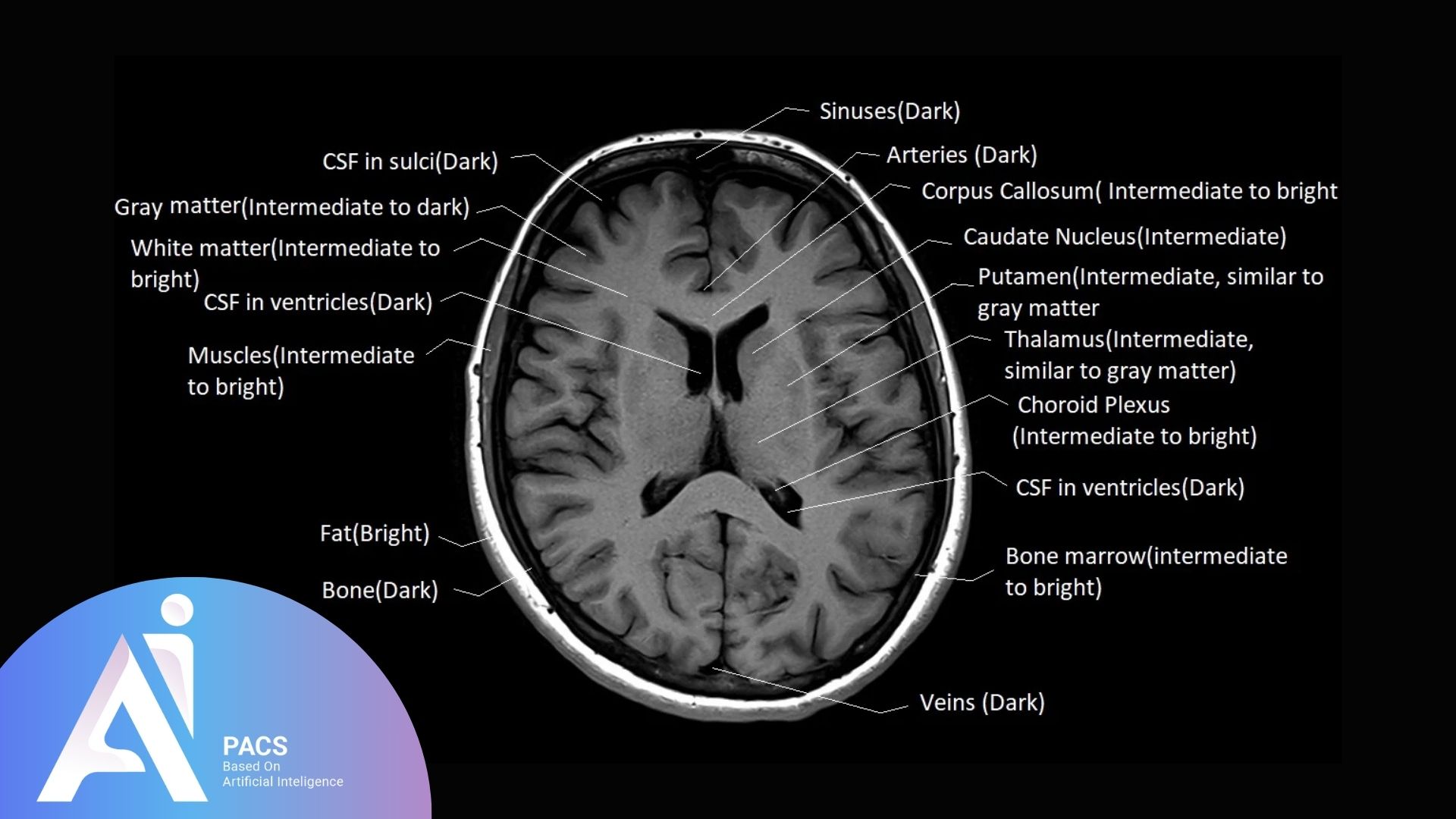
- Hyperintense/Hypointense: Refers to areas on the MRI that appear brighter or darker, respectively, indicating differences in tissue characteristics.
- Enhancement: Refers to areas that absorb contrast dye, often indicating abnormal tissue such as inflammation or tumors.
- Lesion: A general term for any abnormal tissue change, such as a tumor or cyst.
Anatomical Terms and Locations
MRI reports often reference specific anatomical locations. Understanding terms like “anterior” (front), “posterior” (back), “medial” (toward the middle), and “lateral” (toward the side) can help you better interpret the findings.
Imaging Sequences Explained
MRI images are obtained using different sequences, each offering unique insights:
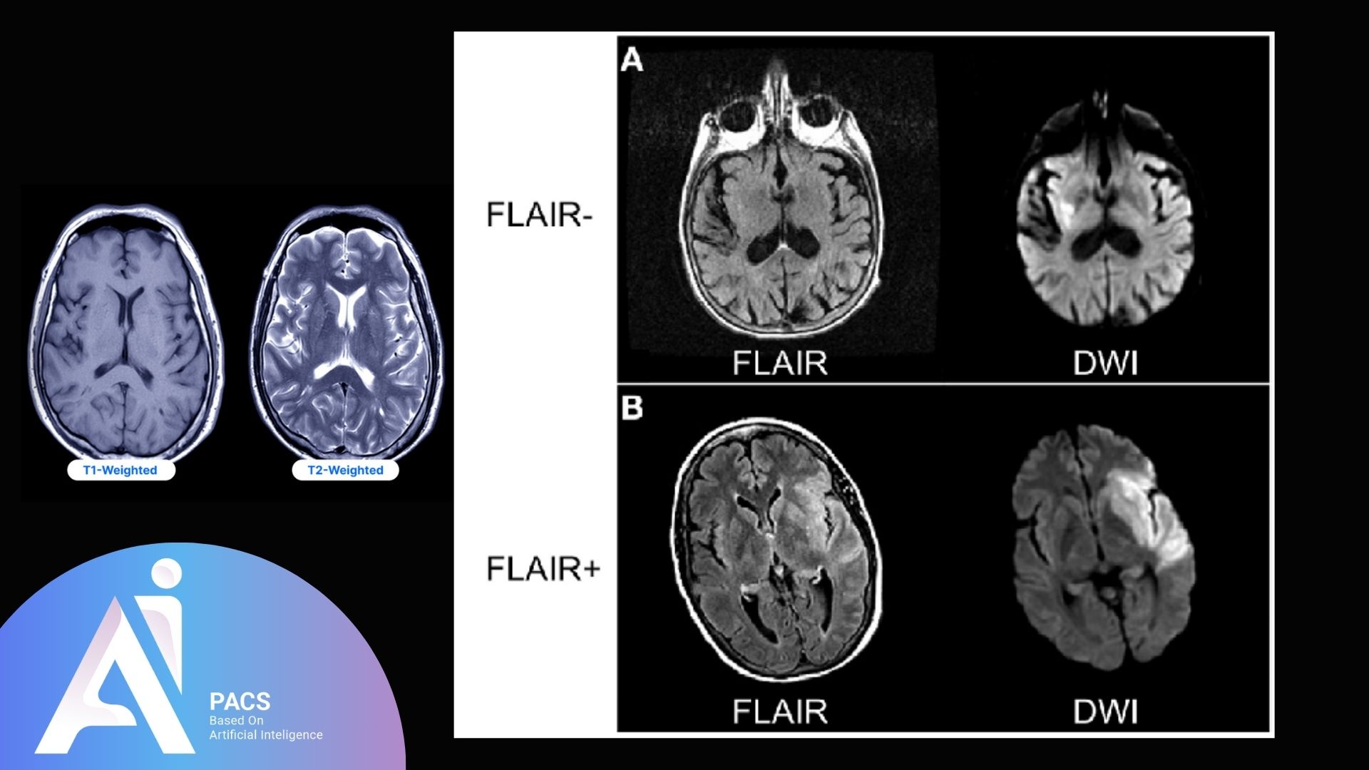
- T1-weighted images: Best for showing the anatomy and structure of organs. Fat appears bright, and water is dark.
- T2-weighted images: Highlight fluids like edema or inflammation, making them helpful in identifying abnormalities.
- FLAIR (Fluid-Attenuated Inversion Recovery): Suppresses normal fluid, making abnormal fluid accumulation, like lesions, more visible.
Basic Structure of an MRI Report
MRI reports typically include several standard sections:
Patient Information Section
This part includes the patient’s name, age, and identification number. It may also note the type of MRI scan performed, such as brain, spine, or joint MRI.
Clinical History
This section provides context for the MRI, explaining why it was ordered, such as for symptoms like headaches or back pain. Understanding this part helps you relate the findings to the reason for the scan.
Description of Findings
The findings section is the report’s core, where the radiologist describes what they see. If any finding remains unclear after you read it, an independent MRI second opinion can confirm the interpretation. It may include descriptions of normal structures as well as any abnormalities observed. For example:
- Brain MRI: May describe the ventricles, gray, and white matter.
- Spine MRI: This test may reveal details about disc conditions, spinal cord alignment, and abnormalities, such as disc bulges.
- Joint MRI: May mention the condition of cartilage, ligaments, and joint fluid.
Impression/Conclusion Section
The impression summarizes the radiologist’s primary observations and is often the most critical part for patients. It may note whether any abnormalities are present and provide a differential diagnosis. This section is key for understanding the overall significance of the findings.
Reading Different Types of MRI Reports
Reading a Brain MRI Report
Brain MRIs may mention:
- Lesions or plaques: Often associated with conditions like multiple sclerosis.
- Atrophy: Indicates shrinkage of brain tissues, possibly related to aging or neurodegenerative diseases.
- Hydrocephalus: Refers to excess fluid in the brain’s ventricles.
Reading a Spine MRI Report
Key terms in spine MRIs include:
- Disc herniation: A common cause of back pain, where the disc protrudes and may compress nerves.
- Spinal stenosis: Narrowing of the spinal canal can cause nerve compression.
- Spondylosis: Degenerative changes in the spine, often due to aging.
Reading a Joint MRI Report
For joint MRIs, look for:
- Meniscus tears: Common in knee MRIs.
- Ligament injuries: Important for understanding stability issues in the joint.
- Arthritis is characterized by changes in cartilage thickness or the development of bone spurs.
👉 Get started with a professional review! Upload your MRI report for an expert second opinion and clear interpretation.
Common Findings in MRI Reports
Degenerative Changes
Degenerative changes, such as osteoarthritis or degenerative disc disease, refer to the age-related wear and tear that occurs over time. Terms like “narrowed disc spaces” or “joint space reduction” indicate this condition.
Inflammatory Findings
MRI reports may describe inflammation as edema (swelling) or synovitis (inflammation of the joint lining).
Report wording varies by body part; for one organ-specific example, see how to understand a prostate MRI report.
Tumors and Masses
The report might use terms like “enhancing mass” to describe potential tumors. Characteristics such as size, shape, and location help guide further diagnosis and treatment.
Tips for Comparing MRI Findings Over Time
When reviewing follow-up MRI scans, focus on the changes between the old and new reports. Pay attention to whether previously noted abnormalities have changed, remained the same, or increased or decreased in severity.
Consulting a Radiologist for Clarification
If specific terms or findings in the report are unclear, consider discussing them directly with a radiologist or your referring physician. They can provide more detailed explanations.
Prefer a quick visual version? See this guide’s key points in about a minute: How to understand MRI findings quickly (visual story).