T1-Weighted MRI Images
T1-weighted MRI images are one of the primary sequences used in magnetic resonance imaging to provide detailed anatomical views of the body. These images are characterized by their ability to differentiate between tissues based on their relaxation times, offering high-contrast visuals that are especially effective for highlighting fat and certain soft tissues. Medical professionals can accurately detect and evaluate various structural abnormalities using these images.
Specific features of T1-weighted images
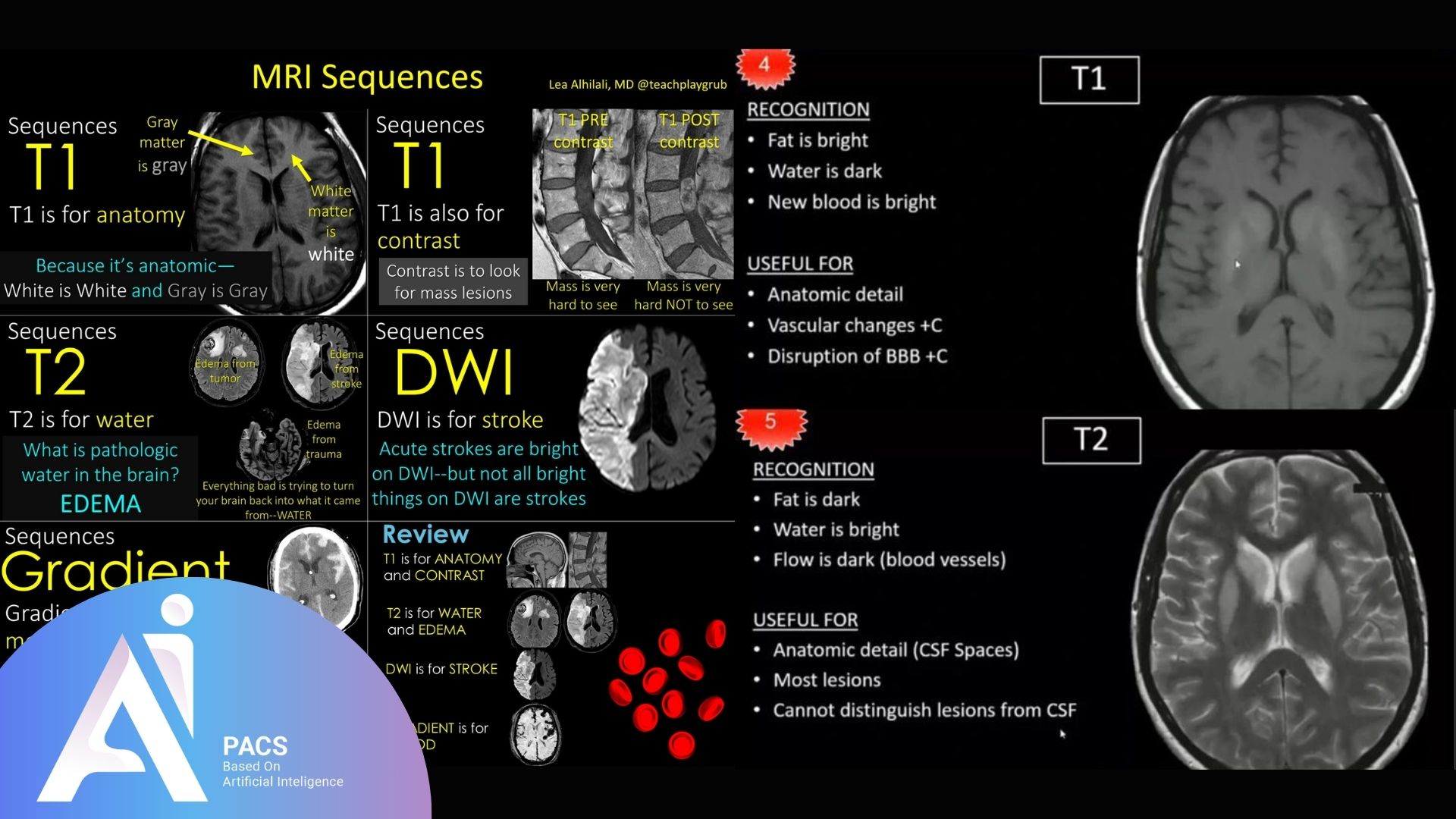
T1-weighted images stand out for their distinct contrast where fat appears bright, and fluids like cerebrospinal fluid (CSF) appear dark. This sequence is particularly valuable for assessing anatomical structures like the brain and musculoskeletal system. Additionally, T1-weighted imaging is often utilized with contrast agents to enhance the visibility of vascular structures and pathological conditions, making it an essential tool in medical diagnostics.
How T1-Weighted MRI Images Work
T1-weighted MRI images are created by manipulating the relaxation properties of protons in tissues. A radiofrequency pulse disrupts this alignment after the magnetic field aligns the protons. T1 imaging measures the time it takes for these protons to realign, or “relax,” with the magnetic field, which varies depending on the tissue type. This relaxation time determines the brightness or darkness of different tissues in the image, allowing for detailed anatomical visualization.
Explanation of tissue contrast and fat signal intensity
In T1-weighted imaging, tissues with shorter relaxation times, such as fat, return to alignment more quickly and appear brighter on the images. Conversely, tissues with longer relaxation times, like fluids (e.g., cerebrospinal fluid), appear darker. This contrast makes T1-weighted images particularly useful for distinguishing between fat, soft tissues, and fluid-filled spaces, offering critical insights into the structural composition of organs and systems.
What T1-Weighted MRI Images Reveal
T1-weighted MRI images provide exceptional clarity in capturing the anatomical details of the body. These images highlight tissue differences, offering valuable insights for diagnosing and monitoring various conditions. They are particularly effective for visualizing structures with high-fat content and are widely used across numerous medical fields, including neurology, orthopedics, and oncology.
High-resolution anatomical details
One of the key strengths of T1-weighted MRI images is their ability to produce high-resolution visuals, making it easier to identify subtle abnormalities. The bright appearance of fat and soft tissue contrasts against darker fluids, providing a clear distinction between different structures. This level of detail is crucial for pinpointing issues like tumors, cysts, or tissue damage, allowing healthcare professionals to make accurate assessments.
Commonly visualized structures
T1-weighted MRI images are commonly used to examine various structures, including the brain, spine, and joints. In brain imaging, they are instrumental in identifying lesions, hemorrhages, and structural anomalies. T1-weighted images help visualize ligaments, tendons, and bone marrow for musculoskeletal evaluations. Additionally, they are widely used in abdominal and pelvic imaging to assess organs like the liver and kidneys, particularly when combined with contrast agents for enhanced diagnostic accuracy.
Applications of T1-Weighted MRI in Diagnosis
T1-weighted MRI images are pivotal in medical diagnostics, offering versatile applications across multiple specialties. Their ability to provide clear contrasts between tissue types makes them indispensable for assessing localized and systemic conditions. Below are the primary diagnostic applications of T1-weighted imaging.
Neurological conditions
T1-weighted imaging is a cornerstone of neurological diagnostics, providing detailed brain and spinal cord visuals. It is frequently used to identify and evaluate tumors, hemorrhages, and structural abnormalities. Post-contrast T1-weighted imaging is particularly valuable for detecting enhancing lesions in multiple sclerosis or metastatic brain tumors. This sequence is also used to assess the integrity of brain structures and diagnose neurodegenerative diseases.
Musculoskeletal evaluations
In orthopedics and sports medicine, T1-weighted MRI images are essential for evaluating soft tissue injuries, bone marrow abnormalities, and joint structures. These images help detect fractures, cartilage damage, and ligament tears with high precision. They are also valuable in identifying bone marrow edema and other subtle changes in musculoskeletal tissues, aiding in early diagnosis and treatment planning.
whole-body imaging and fat distribution
T1-weighted imaging is widely used for whole-body scans, especially when evaluating fat distribution and systemic conditions. This sequence is instrumental in detecting lipomas, assessing fat infiltration in organs like the liver, and monitoring metabolic disorders. Whole-body T1-weighted imaging is also employed in oncology to detect and monitor metastatic lesions, providing a comprehensive view of the body’s internal structures.
Advantages of T1-Weighted MRI Images
T1-weighted MRI images offer several benefits that make them indispensable in medical imaging. These advantages stem from their ability to provide detailed, high-resolution images essential for diagnosing and monitoring various conditions. Below are two key benefits of T1-weighted imaging.
Superior contrast for specific tissues
One of the standout features of T1-weighted MRI is its ability to differentiate tissues with varying fat and water content. Fat appears bright, while cerebrospinal fluid (CSF) appears dark, creating a clear contrast. This makes T1 imaging particularly useful for visualizing fat-rich areas, such as bone marrow, subcutaneous tissues, and specific organs. When used with contrast agents, the imaging further enhances the visualization of abnormal tissue growths, vascular structures, and tumors, offering exceptional diagnostic clarity.
Accuracy in structural imaging
T1-weighted images provide highly accurate depictions of anatomical structures, making them ideal for assessing fine details. Whether identifying subtle abnormalities in the brain, evaluating musculoskeletal injuries, or detecting early signs of disease, T1-weighted imaging captures precise and reliable structural information. This accuracy is crucial for treatment planning and monitoring progress, ensuring better patient outcomes.

Comparison Between T1 and T2-Weighted Images
T1-weighted and T2-weighted MRI images are two foundational sequences in magnetic resonance imaging, each serving unique diagnostic purposes. While they both provide crucial insights, their differences in tissue contrast and applications make them complementary tools in medical imaging. Not sure which MRI type you need? 👉 Get a second opinion on your MRI report today.
Tissue Contrast and Appearance
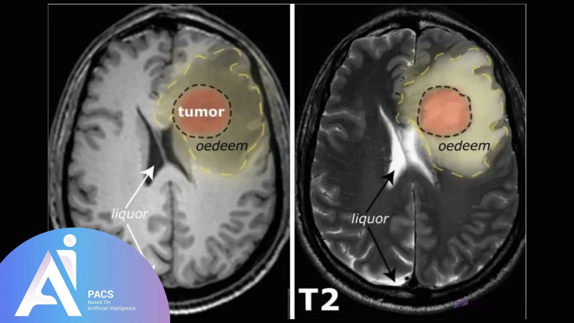
- T1-Weighted Images: Fat appears bright, and fluids like cerebrospinal fluid (CSF) are dark. This makes T1 imaging ideal for highlighting anatomical structures and differentiating between fat and other soft tissues.
- T2-Weighted Images: Fluids are bright, while fat and other tissues appear darker. This high sensitivity to water content makes T2 images suitable for detecting fluid-filled structures, such as cysts, edema, and inflammation.
Diagnostic Applications
- T1-Weighted Images: Used for anatomical detail, T1 images are valuable in evaluating structural abnormalities, fat distribution, and areas enhanced with contrast agents, such as tumors or vascular lesions.
- T2-Weighted Images: Preferred for identifying pathological changes, T2 images excel in detecting inflammation, edema, and fluid accumulation in tissues, making them crucial for diagnosing infections, injuries, and certain chronic conditions.
Strengths and Limitations
- T1-Weighted Images: Offer superior resolution and clarity for structural imaging but are less effective for detecting subtle changes associated with fluid or inflammation.
- T2-Weighted Images: Highly sensitive to pathological changes but may lack the sharp structural detail of T1 imaging.
Complementary Role in Imaging
T1 and T2-weighted images are often used to comprehensively understand a condition’s structural and pathological aspects. This combination helps clinicians make accurate diagnoses and tailor treatment plans to patients’ needs.
Looking for insights on how T2-weighted MRI images assist in identifying brain and spinal cord conditions? Explore this in-depth look at T2-weighted MRI
Conclusion
T1-weighted MRI images are vital in diagnostic imaging, offering excellent clarity for visualizing anatomical structures and detecting abnormalities. They provide superior contrast for fat-rich tissues, making them important for diagnosing neurological conditions, musculoskeletal injuries, and systemic disorders. When combined with T2-weighted imaging, they offer a comprehensive view for accurate diagnosis and treatment planning, playing a crucial role in enhancing medical care and ensuring reliable healthcare diagnostics.


