


")
What Is an AVM, and what does AVM (Arteriovenous Malformation) on MRIs look like?
An arteriovenous malformation (AVM) is a rare and abnormal connection between arteries and veins, bypassing the capillary system. This tangle of blood vessels disrupts the normal blood flow, leading to potential complications in the affected area. AVMs can occur anywhere in the body but are most commonly found in the brain and spinal cord. While some AVMs are present from birth, they may not cause symptoms until later in life, depending on their size and location.
How it affects blood flow and surrounding tissues
In a healthy vascular system, arteries carry oxygen-rich blood from the heart to tissues, and veins return oxygen-depleted blood to the heart. This process is disrupted in an AVM because blood flows directly from arteries to veins without passing through capillaries. This can lead to:
- Inadequate oxygen delivery: Nearby tissues may suffer from oxygen deprivation, leading to cell damage.
- Increased pressure on veins: The abnormal blood flow can weaken veins, making them prone to rupture.
- Potential for hemorrhage: In the brain, this can result in life-threatening conditions like stroke or seizures.
Over time, the strain on surrounding tissues can cause further complications, emphasizing the importance of early detection and management.
Common Symptoms of AVM
Arteriovenous malformations (AVMs) can present with various symptoms depending on their size, location, and the extent of the disruption they cause to normal blood flow. In many cases, AVMs are discovered only when complications arise, such as bleeding or significant neurological issues. Early recognition of symptoms is critical for timely diagnosis and management.
Neurological symptoms and warning signs
When AVMs occur in the brain or spinal cord, they often manifest with neurological symptoms, including:
- Headaches: Persistent or severe headaches, sometimes resembling migraines.
- Seizures: Sudden, unexplained seizures are a common first symptom of a brain AVM.
- Weakness or numbness: Unilateral weakness or loss of sensation, particularly in the limbs.
- Vision disturbances: Blurred vision, partial vision loss, or other visual impairments.
- Difficulty speaking or understanding speech: These symptoms can mimic a stroke.
These symptoms often occur suddenly and may indicate an underlying AVM that requires urgent medical attention.
How AVM can present in asymptomatic cases
Not all AVMs cause noticeable symptoms. Many cases remain asymptomatic for years and are often found incidentally during imaging for unrelated issues. Factors contributing to asymptomatic cases include:
- Small AVM size: Smaller AVMs may not disrupt surrounding tissues significantly.
- Stable blood flow: Some AVMs do not cause pressure buildup or oxygen deprivation in nearby tissues.
- No associated bleeding: The absence of rupture or hemorrhage minimizes overt symptoms.
While asymptomatic AVMs may seem harmless, they still pose a risk of future complications, such as sudden bleeding or neurological damage. Regular monitoring through imaging is crucial for managing these silent cases.
")
Role of MRI in Diagnosing AVM
MRI (Magnetic Resonance Imaging) is crucial for diagnosing arteriovenous malformations (AVMs). It offers a detailed visualization of the abnormal blood vessels and surrounding structures. Its ability to provide high-resolution images without radiation makes it the preferred choice for both initial detection and ongoing monitoring of AVMs.
Why MRI is the gold standard for AVM detection
MRI is considered the gold standard for diagnosing AVMs due to several advantages:
- Detailed imaging: MRI provides precise, high-resolution images of soft tissues and blood vessels, enabling accurate identification of AVMs.
- Non-invasive and radiation-free: Unlike CT scans, MRI avoids radiation exposure, making it safer for repeated evaluations.
- Multiple imaging modes: Techniques like MR angiography (MRA) enhance blood flow and vessel abnormalities visualization.
- Early detection of complications: MRI can identify signs of bleeding, surrounding tissue damage, or edema, even in small or asymptomatic AVMs.
These features make MRI indispensable in diagnosing AVMs and assessing their severity for treatment planning.
Critical features of AVM visible on MRI
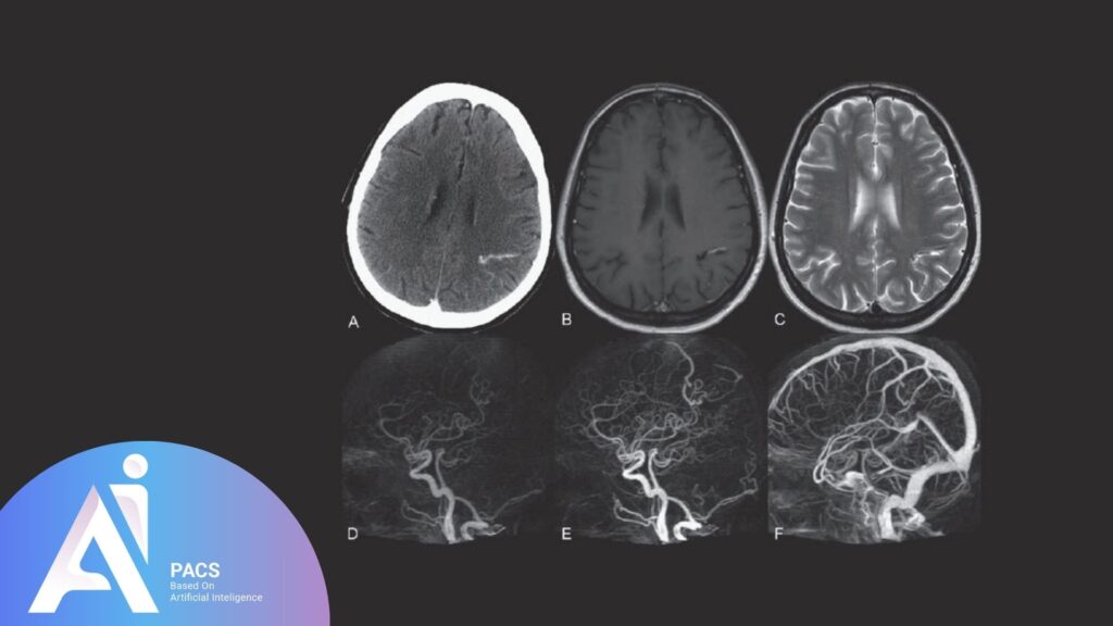
When imaging an AVM, MRI reveals several distinct characteristics that help in diagnosis and evaluation:
- Tangled blood vessels: The hallmark feature of AVMs is a cluster of abnormal blood vessels visible on contrast-enhanced MRI or MRA.
- Absence of capillary network: The lack of a normal capillary bed is evident in the images.
- Feeding arteries and draining veins: MRI can delineate the arteries supplying the AVM and the veins draining it, providing crucial insights for treatment planning.
- Signs of hemorrhage: Areas of bleeding appear as dark or bright spots, depending on the imaging sequence used.
- Surrounding tissue changes: Swelling, edema, or ischemia in nearby tissues can also be visualized.
These features make MRI an essential tool for clinicians in diagnosing, monitoring, and managing AVMs effectively.
Types of MRI Techniques Used for AVM
MRI offers techniques that provide detailed insights into arteriovenous malformations (AVMs). Each method highlights specific aspects of the AVM, from structural abnormalities to functional dynamics, aiding in comprehensive diagnosis and treatment planning.
Standard MRI vs. MR angiography (MRA)
Standard MRI:
This technique uses high-resolution brain or spinal cord imaging to identify AVM-related structural changes. It is beneficial for:
- Detecting the tangled mass of blood vessels characteristic of AVMs.
- Identifying complications such as edema, ischemia, or hemorrhage in surrounding tissues.
- Providing an initial assessment of AVM size and location.
MR Angiography (MRA):
MRA is a specialized MRI technique designed to visualize blood flow within vessels. It is often used in AVM cases to:
- Map feeding arteries and draining veins.
- Assess the extent of vascular abnormalities.
- Provide a non-invasive alternative to traditional angiography for vascular imaging.
By combining standard MRI with MRA, clinicians can comprehensively view the AVM’s dranaging and vascular aspects.
Functional imaging and contrast-enhanced scans
Functional MRI (fMRI):
This technique measures brain activity by detecting changes in blood flow. It is precious for AVMs located in critical brain regions, helping to:
- Determine how the AVM affects surrounding functional areas.
- Plan surgical or radiological interventions to minimize neurological impact.
Contrast-Enhanced MRI:
Contrast agents enhance the visibility of blood vessels and surrounding tissues. This method helps:
- Highlight the AVM structure and its vascular network.
- Differentiate between feeding arteries, draining veins, and regular vessels.
- Detect subtle changes in blood flow patterns and signs of inflammation or bleeding.
These advanced MRI techniques ensure precise diagnosis, enabling tailored treatment strategies for AVM management.
Interpreting MRI Results for AVM
Understanding the MRI results for an AVM (arteriovenous malformation) is critical in determining its severity, location, and impact on surrounding tissues. These interpretations guide treatment decisions and help predict potential complications. Below are the key factors clinicians evaluate when interpreting MRI results for AVMs.
”
Ident”fying the AVM Location and Size
Location: The MRI pinpoints the exact location of the AVM, which is crucial in assessing its risk. AVMs in the brainstem or spinal cord are particularly concerning due to the potential for severe neurological complications.
Size: The size of the AVM is a critical factor; larger AVMs are generally more likely to cause symptoms like bleeding or pressure effects on nearby tissues.
Examining Feeding Arteries and Draining Veins
MRI, especially when combined with MR angiography (MRA), allows detailed visualization of the AVM’s vasAVM’s structure:
- Feeding arteries: Identifies the arteries supplying blood to the AVM.
- Draining veins: This map shows the veins carrying blood away from the AVM. It helps assess the flow dynamics and the risk of rupture.
Assessing Signs of Hemorrhage or Tissue Damage
Bleeding: Evidence of acute or past bleeding is often visible as dark or bright spots, depending on the MRI sequence used. This is a critical indicator of an AVM’s risk.
Ischemia and Edema: MRI shows signs of oxygen deprivation or swelling in tissues surrounding the AVM, which can contribute to symptoms like headaches or neurological deficits.
Evaluating Surrounding Structures
MRI helps assess how the AVM interacts with nearby structures, such as the brain or spinal cord, to determine whether it is causing compression, inflammation, or disruption of normal function.
Interpreting MRI findings precisely is essential for developing a comprehensive treatment plan, whether it involves surgery, embolization, radiosurgery, or conservative monitoring. Regular MRI follow-ups are often necessary to track changes in the AVM over time.
Treatment Planning with MRI Findings
MRI findings are essential for planning treatment for AVM (arteriovenous malformations). They provide critical information about the AVM’s size, location, and vascular structure, aiding in the decision to intervene and guiding treatment options like surgery or embolization. When combined with MR angiography (MRA), MRI helps identify feeding arteries and draining veins for precise targeting during procedures. It also assesses surrounding tissue damage and complications, which aids in risk evaluation. Regular MRI monitoring is vital for managing AVMs and detecting recurrence after treatment.
Risks and Limitations of MRI for AVM
While MRI is a powerful tool for diagnosing and evaluating AVMs, it does have some risks and limitations that must be considered:
Risks Associated with MRI
- Claustrophobia: The enclosed nature of MRI machines can cause anxiety or discomfort in some patients.
- Contrast agent reactions: Some individuals may experience mild allergic reactions or, in rare cases, kidney complications during contrast-enhanced MRI, particularly in those with pre-existing kidney issues.
- Metal implants or devices: Due to safety concerns, patients with certain types of metal implants, pacemakers, or other medical devices may not be eligible for MRI.
Limitations of MRI for AVM
- Small or asymptomatic AVMs: Tiny AVMs or those without significant blood flow abnormalities may be challenging to detect, especially on standard MRI without advanced techniques like MR angiography.
- Motion artifacts: Patient movement during the scan can reduce image quality, making it difficult to identify and evaluate the AVM accurately.
- Cost and availability: MRI is expensive and may not be readily available in all healthcare settings, limiting access for some patients.
- Lack of functional assessment: While MRI provides detailed structural information, it may not fully capture the functional impact of the AVM, such as its effect on blood flow or surrounding neural activity, without additional specialized imaging like functional MRI (fMRI).
Despite these challenges, MRI remains the gold standard for AVM diagnosis, and its limitations can often be mitigated with complementary imaging techniques and proper patient preparation.
Conclusion
In conclusion, MRI is a vital tool for diagnosing and managing arteriovenous malformations (AVMs). It provides detailed visualization of their structure, size, and impact on surrounding tissues. Advanced techniques like MR angiography allow for precise assessments that are critical for treatment planning.
Despite some limitations, such as difficulty detecting small AVMs and contraindications for certain patients, the benefits of MRI far outweigh these drawbacks. MRI significantly improves outcomes for individuals with AVMs by enabling early detection and guiding appropriate interventions.