


Understanding Your Breast MRI Report: What the Results Really Tell You
Understanding your breast MRI report is crucial. It may include technical terms such as FGT, BPE, or non-mass enhancement. These words can be confusing, especially when you’re trying to understand whether your results are normal or concerning. But with the right knowledge, you can take control of your health journey.
This guide will help you understand the most common terms found in breast MRI reports, especially if the imaging follows the North American BI-RADS reporting structure.
About the Author
I’m Dr. Vahid Alizadeh. In this article from the “What Does My Report Say?” series, I’ll explain the common terminology used in breast MRI reports, enabling you to understand your results and next steps better.
👉 Upload Your Breast MRI Report for Expert Review
Real-Life Scenario: A Patient’s Journey
Jessica, a 41-year-old woman, had a strong family history of breast cancer and underwent a screening breast MRI. Her report included phrases like fibroglandular tissue, BPE, and segmental non-mass enhancement. Not sure if she should be worried, she turned to us for a clear explanation.
What Do These Medical Terms Mean?
FGT – Fibroglandular Tissue
The part of the breast composed of fibrous and glandular tissue, not fat.
Why It Matters: The report will describe the extent of FGT present. A higher amount can make it more difficult to detect abnormalities.
Background: The breast is made of two key tissue types—fat and fibroglandular tissue. The glandular part is responsible for milk production.
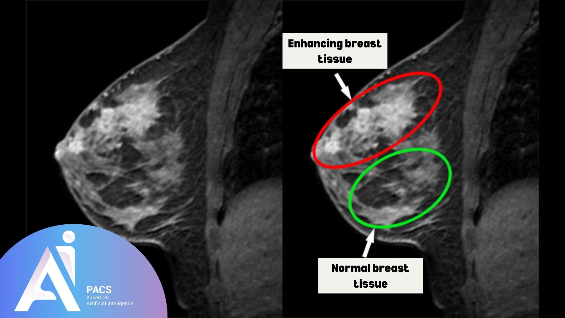
BPE – Background Parenchymal Enhancement
The normal uptake of contrast in breast tissue after contrast injection.
Why It Matters: High BPE can obscure underlying lesions and reduce the MRI’s sensitivity.
Tip: Breast MRIs are often scheduled in the early follicular phase of the menstrual cycle (days 7–14) to minimize BPE.
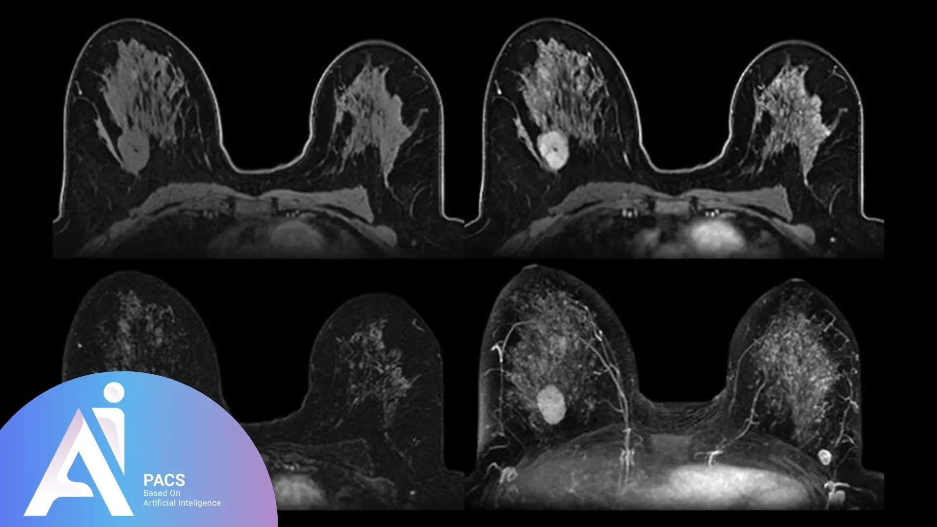
Mass Description
If a mass is seen, the report will describe:
- Shape: Round, oval, irregular
- Margins: Circumscribed (well-defined) or spiculated (irregular)
- Size and location: Dimensions and quadrant of the breast
Why It Matters: These characteristics help determine whether a lesion is likely benign or suspicious.
Non-Mass Enhancement (NME)
Areas of abnormal contrast uptake that don’t form a distinct mass.
Why It Matters: NMEs can appear in patterns that may raise concern depending on their shape and distribution.
Common Descriptors of NME
- Clumped: Patchy, clustered appearance; may raise suspicion
- Heterogeneous: Mixed intensity; nonspecific but may require follow-up
- Linear: Follows ducts or vessels; can be benign or suspicious
Distribution Patterns of NME
- Segmental: Follows one ductal system; potentially worrisome
- Regional: Covers a large area but not a ductal pattern
- Focal: Small, well-defined area
- Diffuse: Spread throughout the breast
Why It Matters: Segmental and clumped patterns are more concerning and may require further testing.
BI-RADS Assessment Category
The BI-RADS (Breast Imaging Reporting and Data System) is a standardized system used in North America to classify breast imaging findings. Categories include:
- BI-RADS 1: Negative
- BI-RADS 2: Benign findings
- BI-RADS 3: Probably benign – short-term follow-up recommended
- BI-RADS 4: Suspicious abnormality – biopsy should be considered
- BI-RADS 5: Highly suggestive of malignancy – appropriate action should be taken
- BI-RADS 6: Known biopsy-proven cancer – imaging for treatment planning or monitoring
What Does This Report Say About My Condition?
Understanding the type of enhancement, the amount of fibroglandular tissue, and how findings are categorized under BI-RADS can help guide your next steps. For example:
- High BPE might explain why follow-up is recommended despite no clear mass.
- A BI-RADS 3 or 4 suggests the need for monitoring or biopsy.
- A clumped, segmental, non-mass enhancement may require additional imaging.
Why You Should Get an Expert to Review Your Report
Breast MRI interpretation is complex. An expert radiologist can:
- Distinguish between benign and suspicious patterns
- Determine if follow-up or biopsy is needed
- Help you understand the BI-RADS category in your context
At AI-PACS, our breast imaging experts provide second opinions to ensure that nothing important is overlooked. Getting a second opinion can provide you with peace of mind and ensure that you receive the best possible care.
Next Steps: Let’s Help You Understand Your Report
If you’ve received a breast MRI report and aren’t sure what the findings mean, upload it for expert interpretation.
👉 Upload your report now and get an expert review from our online radiology report services.
“If you’re unfamiliar with how breast MRI works, check out our full guide to the procedure here.”
Final Thought
Understanding your breast MRI report is an essential step in taking control of your health. While medical terminology can seem overwhelming, breaking down terms like FGT, BPE, and non-mass enhancement helps demystify the process and provides clarity on what’s really important. Remember, your health is your most valuable asset, and having an expert review your results can offer peace of mind and guide you toward the right next steps. If you’re ever in doubt, don’t hesitate to seek a second opinion from a specialist to ensure you’re making informed decisions about your care.