


Critical Reasons to Get Imaging for Back Pain
Back pain is a common condition that affects millions of people worldwide and can significantly impact daily life and productivity. It can range from mild, temporary discomfort to chronic pain that severely limits daily activities and overall quality of life. While many cases are self-limiting and respond well to rest, physical therapy, or over-the-counter medication, others may persist or worsen over time. In such cases, back pain could be a sign of more serious underlying issues, such as disc herniation, spinal degeneration, or even infection or cancer. Imaging tests like MRIs, CT scans, and X-rays play a crucial role in identifying these hidden problems early, enabling timely and targeted treatment before complications arise.
If you’re seeking reliable online radiology report services, AI-PACS is here to provide you with the best second opinion on your medical images, delivered by our team of expert radiologists.
Common Causes of Back Pain
Back pain can arise from numerous causes, each requiring specific diagnostic and therapeutic approaches. Common origins include:
1. Discopathies and Disc Herniations
- Intervertebral discs are the soft, cushion-like structures that sit between the bones (vertebrae) in your spine. Over time, these discs can become damaged or degenerate. A herniated disc, also called a slipped or ruptured disc, occurs when the soft center of the disc pushes through a tear in the outer layer.
- A herniated disc can put pressure on nearby nerves, leading to nerve compression. This results in symptoms like sharp or shooting pain, numbness, or weakness in the affected area, often radiating to the legs or arms.
- Imagine you’re bending over to pick something up, and you feel a sharp pain shoot down your leg. This could be a sign of a herniated disc pressing on the sciatic nerve, causing sciatic pain, or “sciatica.”
2. Osteoarthritis and Degenerative Changes
- Osteoarthritis (OA) is a progressive condition where the protective cartilage that cushions the joints breaks down. In the spine, this can affect the facet joints (the joints that allow the spine to move), leading to inflammation, stiffness, and pain.
- As the cartilage wears down, bones begin to rub together, which causes pain, swelling, and limited mobility. Over time, OA can also lead to bone spurs (osteophytes) that can impinge on nearby nerves.
- You may notice stiffness or aching in your lower back, especially after sitting for long periods or when getting out of bed in the morning. This could indicate that OA is affecting the joints in your spine.
3. Facet Joint Injuries and Posterior Element Damage
- The facet joints are the small joints located at the back of the spine. These joints allow for the smooth movement of your spine as you bend and twist. When these joints are injured, it can lead to localized pain and inflammation.
- Facet joint injuries typically occur due to trauma or repetitive strain, leading to inflammation and irritation in the joints. The pain is often felt in the lower back and can worsen with movement or prolonged standing.
- A car accident that causes whiplash can result in a facet joint injury. You may experience pain that is localized to the lower back or neck, often aggravated by twisting or bending.
4. Traumatic Injuries, Including Vertebral Fractures
- Vertebral fractures occur when a bone in the spine cracks or breaks, often due to trauma or injury. Common causes include accidents, falls, or sports-related injuries.
- A vertebral fracture can cause immediate, intense pain in the back. It may also result in spinal instability, leading to longer-term issues if not treated properly.
- A person who falls from a height or is involved in a car accident may experience severe back pain and difficulty moving. A vertebral fracture could be the cause, and medical imaging such as an X-ray or MRI would be required to confirm the diagnosis.
5. Pathological Fractures (e.g., Metastatic Cancer)
- A pathological fracture occurs when a bone becomes weakened by an underlying condition, such as osteoporosis or cancer. In cases of metastatic cancer, cancer from other parts of the body spreads (metastasizes) to the spine, weakening the bone.
- The weakened bone is more prone to fractures with minimal trauma or everyday activities. These fractures can cause significant pain, especially if the vertebrae collapse and press on nearby nerves.
- A patient with a history of breast cancer may experience new-onset back pain. If the cancer has spread to the spine, it could lead to a pathological fracture, causing severe discomfort, especially with movement.
6. Infectious Processes, Such as Osteodiscitis
- Osteodiscitis is an infection that affects the vertebrae or the intervertebral discs. It can result from a bacterial infection, often following surgery, trauma, or a bloodstream infection that spreads to the spine.
- The infection leads to inflammation and swelling in the spine, which can cause intense, localized pain, often accompanied by fever or other signs of infection. If left untreated, it can lead to further complications.
- A person who has recently undergone spinal surgery or has a history of infections may develop symptoms like a fever, persistent back pain, and tenderness in the spine. If this is suspected, imaging and lab tests are essential to confirm the infection.
7. Inflammatory Conditions
- Conditions like ankylosing spondylitis (AS), rheumatoid arthritis, and other autoimmune diseases cause chronic inflammation in the spine and other joints, leading to pain, stiffness, and reduced mobility.
- Inflammatory conditions often affect the sacroiliac joints and other parts of the spine. Over time, the inflammation can cause joint fusion (in AS) or severe damage to the joints, resulting in chronic pain.
- A person with ankylosing spondylitis may experience lower back pain that worsens at night and improves with movement. Over time, the inflammation can lead to stiffness and difficulty bending.

Recognizing Red Flags in Back Pain
Recognizing “red flag” symptoms is crucial, as they indicate potentially serious conditions that require urgent evaluation. These symptoms include:
1. Fever or Signs of Infection
- Fever, chills, night sweats, or abnormal blood test results (such as elevated inflammatory markers) can indicate an infection in the spine or nearby structures.
- Infections like osteomyelitis (bone infection) or osteodiscitis (infection of the intervertebral discs) can cause severe spinal discomfort. If left untreated, infections can spread to other areas of the body, leading to life-threatening complications.
- Imagine you have back strain that worsens over several days. Along with this discomfort, you start experiencing chills, a fever, and night sweats. These are all signs that an infection may be present in your spine, and immediate medical evaluation, including imaging, is necessary to confirm the diagnosis and begin treatment.
2. Localized Spinal Tenderness
- Significant, localized pain or tenderness when pressure is applied to a specific area of the spine can be a sign of a fracture or localized infection.
- This symptom often suggests that a specific area of the spine is injured or infected. Fractures caused by trauma or pathological conditions (like cancer) can be easily overlooked without proper imaging.
- After lifting a heavy object, you notice sharp pain when pressing on your lower back. The tenderness is specific to one spot, and it doesn’t improve with typical treatments like rest or over-the-counter pain relievers. This could indicate a fracture or an infection that requires further imaging (such as an MRI or X-ray) to diagnose accurately.
3. Neurological Deficits
- Symptoms such as muscle weakness, loss of sensation, numbness, altered reflexes, or difficulty walking may indicate nerve compression or damage.
- These symptoms can result from conditions such as a herniated disc, spinal stenosis, or a tumor pressing on the spinal cord or nerve roots. If left unaddressed, nerve damage can lead to permanent disability.
- You’re experiencing persistent spine-related pain, but you also begin to notice that your leg feels weak, and you have difficulty walking or standing. Your foot may feel “numb,” and you notice a change in your reflexes. These neurological symptoms point to possible nerve compression, and urgent imaging is needed to assess the underlying cause.
4. Recent Significant Trauma
- Any recent fall, accident, or direct injury to the back or spine is an important red flag, especially if it leads to new or worsening pain.
- Trauma, such as a fall or car accident, can result in spinal fractures, dislocations, or soft tissue injuries that may not be immediately obvious. Delayed treatment of these injuries can lead to long-term problems, including spinal instability.
- After falling from a height, you start experiencing severe lower back issues that don’t improve with rest. You might also feel a sudden loss of mobility. Even if you didn’t immediately feel the extent of the injury, imaging is crucial to rule out fractures or other spinal injuries.
5. Unexplained Weight Loss or Known History of Cancer
- Unexplained weight loss, particularly in combination with back pain, could suggest that the pain is related to a systemic disease such as cancer. If you have a history of cancer, this could be a sign that the cancer has spread (metastasized) to the spine.
- Certain types of cancer, such as breast, lung, or prostate cancer, can spread to the spine. Metastatic cancer can cause structural changes in the spine and lead to fractures, nerve compression, and other complications.
- A person with a history of breast cancer begins to experience persistent spinal discomfort and notices unexplained weight loss. This could be an indication that the cancer has spread to the spine, and urgent imaging, such as an MRI or CT scan, is needed to confirm the diagnosis and plan appropriate treatment.
6. Age Greater Than 50 with New-Onset Severe Pain
- If you’re over 50 and experience sudden, severe backache, it raises concerns about conditions like vertebral fractures, infections, or malignancies.
- Older individuals are more susceptible to conditions such as osteoporosis (which can lead to fractures) or spinal infections. Additionally, the risk of cancers, such as metastatic tumors, increases with age.
- A 60-year-old starts experiencing severe, sudden lower back pain after bending over. The pain does not improve with rest and feels different from usual muscle strain. This could be a sign of a vertebral fracture due to osteoporosis or other serious conditions, and imaging is needed to rule out underlying issues.
7. Night or Rest Pain
- Backache that worsens at night or during periods of rest can indicate that something more serious is happening, such as an infection, tumor, or inflammatory condition.
- Pain that intensifies at night or when lying down may be a sign of cancer, infection, or inflammation, all of which require urgent evaluation. Unlike typical muscle strain, this type of pain does not improve with movement or activity.
- You notice that your spinal distress becomes worse at night, keeping you awake or disturbing your sleep. You’ve also tried resting, but the pain persists or even intensifies. This could indicate a deeper issue, like an infection or a tumor pressing on the spine, and imaging should be done to investigate further.
Urgent Imaging is Essential
When any of these red flag symptoms appear, timely imaging is crucial to ensure an accurate diagnosis and appropriate treatment. Diagnostic tests such as X-rays, MRIs, or CT scans can identify the root cause of the pain and help doctors decide on the most effective treatment plan. Early diagnosis of serious conditions can prevent further complications, minimize damage, and improve the prognosis.
If you experience any of these red flags along with spinal discomfort, it’s important to consult with a healthcare professional as soon as possible for the appropriate imaging and evaluation.
The Crucial Role of Imaging in Diagnosing Back Pain
Differentiating Causes of Pain
Imaging is essential in accurately distinguishing between various potential causes of back pain:
Discopathies (Disc Herniations and Degeneration):
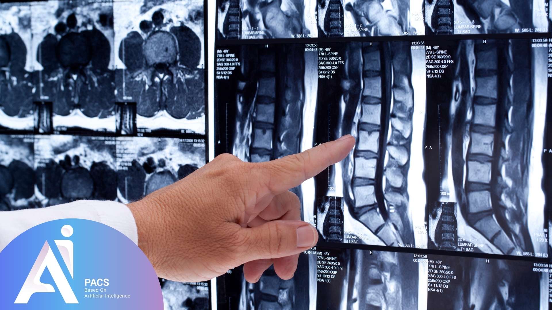
One of the most common causes of lumbar pain is disc-related problems. An MRI (Magnetic Resonance Imaging) scan provides detailed images of the intervertebral discs, which can reveal herniations (when the disc bulges out and presses on nearby nerves) and degeneration (when the disc wears down). For instance, a person experiencing radiating pain down their leg may have a herniated disc compressing the sciatic nerve, and an MRI would show the precise location and severity of the disc bulge. This helps guide decisions regarding treatment, such as physical therapy, injections, or surgery.
Osteoarthritis and Degenerative Changes:
As we age, the cartilage in our spinal joints can wear down, leading to osteoarthritis. X-rays and MRIs are critical in assessing the extent of joint deterioration and the formation of bone spurs (osteophytes). For example, someone with chronic back stiffness and discomfort after sitting for long periods might be experiencing osteoarthritis in the spine. Imaging will show the level of joint damage and help determine whether conservative treatments, like anti-inflammatory medications or physical therapy, will suffice, or if more advanced treatments, like injections or surgery, are necessary.
Traumatic Injuries:
Trauma to the spine—whether from a fall, sports injury, or accident—can result in fractures, dislocations, or soft tissue injuries. Imaging allows for a clear assessment of the extent of the injury. For instance, a person who has fallen and is experiencing significant spinal discomfort may have a vertebral fracture, and an X-ray or CT scan can immediately detect fractures or dislocations, facilitating quicker diagnosis and treatment.
Pathological Fractures:
Pathological fractures are fractures that occur in bones weakened by underlying diseases such as cancer (metastatic cancer) or osteoporosis. Advanced radiological assessments, particularly CT scans and MRIs, help in detecting these fractures, which may not be visible on X-rays. For example, a person with a known history of lung cancer who suddenly develops new back pain may be dealing with metastatic cancer in the spine. Diagnostic tests will be necessary to confirm the spread of the cancer and plan the appropriate treatment.
Understanding the Standard CT Scan Report Format
In many cases, CT scans play a key role in accurately diagnosing the cause of back pain. However, performing the scan alone isn’t enough—the way the images are reported and interpreted is just as important. Understanding the standard format of a CT scan report can help both patients and healthcare providers better comprehend the findings and communicate more effectively about treatment options.
Infectious Processes:
Infections in the spine, such as osteomyelitis or osteodiscitis (infection of the bones or intervertebral discs), can lead to severe back pain that doesn’t improve with typical treatments. Medical imaging is particularly effective in visualizing infections, as it can show inflammation and infection in the vertebrae or discs. A patient with fever, back pain, and signs of infection may have osteodiscitis, and MRI would provide the clear images needed for diagnosis and to guide appropriate antibiotic or surgical intervention.
Inflammatory Conditions:
Inflammatory conditions, such as ankylosing spondylitis or rheumatoid arthritis, can cause chronic inflammation of the spine and joints. Diagnostic scans are valuable in detecting early signs of inflammation and changes in the spinal joints, which may lead to stiffness, pain, and loss of movement. A person with progressive back stiffness, especially in the morning, may have ankylosing spondylitis, and MRI can detect early spinal inflammation, enabling early intervention to prevent long-term damage.


Assessing Severity and Surgical Indications
While clinical assessments and electromyography (EMG) studies remain foundational in treatment planning, imaging plays a significant role in showing the severity of structural abnormalities. Conditions particularly highlighted by imaging include:
Severe Spinal Canal Stenosis:
This condition occurs when the spinal canal narrows, putting pressure on the spinal cord and nerves. Medical imaging, particularly MRI, helps to quantify the degree of narrowing and nerve compression. For example, a person with difficulty walking, leg weakness, and numbness may have severe spinal stenosis, which MRI will clearly reveal, helping guide surgical decisions to relieve pressure on the spinal cord.
Significant Loss of Vertebral Height:
A loss of vertebral height can suggest compression fractures or severe degenerative changes, both of which require immediate attention. X-rays and CT scans can help assess the degree of vertebral collapse. For instance, if a patient experiences sudden, intense back pain after bending over, the loss of vertebral height could indicate a fracture due to osteoporosis, and diagnostic tests would be needed to plan a treatment approach.
Disrupted Spinal Alignment:
Spinal misalignment can lead to chronic pain, instability, and difficulty moving. Spinal evaluations such as X-rays and MRIs can visualize disruptions in the spine’s alignment, which may require surgery to correct. For example, scoliosis or kyphosis (abnormal curvatures of the spine) can be clearly seen on scans, providing valuable information for surgical planning.
Spinal Instability:
Imaging can reveal abnormal spinal mobility that may require stabilization. For example, if a person’s back pain is caused by a condition such as spondylolisthesis, where one vertebra slips over another, CT scans and MRIs will show the degree of instability, guiding surgical decisions if necessary.
Extensive Degenerative Disc Disease:
In cases of advanced disc degeneration, imaging helps determine which specific spinal levels require surgical intervention. MRI is particularly useful for this purpose, as it shows detailed images of the discs and their relationship with surrounding structures. A person with multiple levels of degenerative disc disease may require surgery at specific levels, as seen on MRI, to alleviate pain and improve function.
The Importance of Integrating Diagnostic Scans with Clinical Evaluation
It’s important to note that radiological assessments alone may not always align perfectly with a patient’s clinical symptoms. Scans can identify structural issues, but how those issues relate to the patient’s experience of pain must be evaluated in the context of their clinical history, physical examination, and pain pattern description. For example, some people may have severe disc degeneration visible on an MRI but experience only mild symptoms, while others may have minimal visible issues but suffer from significant pain. Therefore, integrating radiological exams with the patient’s history and a detailed physical examination is essential to formulate the most accurate diagnosis and create an effective treatment plan.
Why a Second Opinion on Imaging is Crucial
Most radiologists primarily interpret MRI or CT scans based solely on imaging findings without correlating them with the patient’s clinical condition. This practice may lead to incomplete or incorrect diagnoses. A second opinion, which carefully considers clinical history, physical examinations, and imaging studies together, provides substantial benefits:
- Confirming or revising initial diagnoses: Ensures diagnostic accuracy by integrating imaging with clinical symptoms and signs.
- Clarifying treatment decisions: Guides effective, patient-specific management strategies tailored precisely to clinical presentations.
- Providing reassurance and confidence: Offers peace of mind through confirmed diagnoses and well-integrated treatment recommendations.
- Avoiding unnecessary or incorrect treatments: Minimizes the risk of ineffective procedures resulting from misinterpreted images.
- Enhanced patient understanding: Patients gain better insight into their condition, helping them actively participate in treatment decisions.
For a detailed, clinically integrated expert review of your imaging studies, obtain a professional second opinion here. To gain further detailed insights and comprehensive information on back pain management, diagnosis, and treatment, visit our comprehensive blog for additional articles and resources. Empower yourself with accurate information to make informed decisions about your spinal health with confidence.
Final Insights
Back pain can stem from a variety of causes, some of which may be serious and require prompt medical attention. Imaging plays a vital role in uncovering the root cause, guiding treatment, and preventing long-term complications. However, accurate diagnosis doesn’t rely on imaging alone—it requires a comprehensive approach that includes clinical evaluation and, when necessary, a second opinion. By staying informed and proactive, patients can take control of their spine health and ensure they receive the most effective, individualized care possible.
references: