


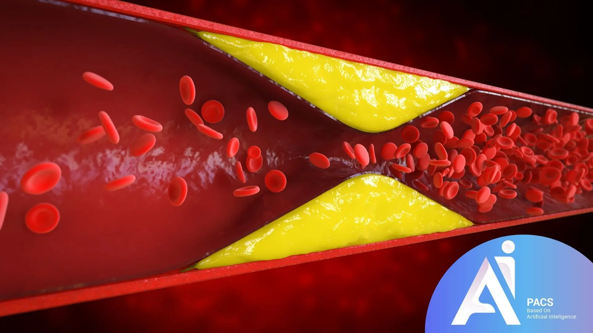
What is Deep Vein Thrombosis (DVT)?
Deep Vein Thrombosis (DVT) is a medical condition where a blood clot (thrombus) forms in the deep veins, usually in the legs. This condition can obstruct blood flow, leading to swelling, pain, and discoloration in the affected area. DVT is a serious health concern because the clot can dislodge and travel to the lungs, causing a potentially life-threatening condition known as a pulmonary embolism (PE). Understanding the causes, symptoms, and risk factors of DVT is crucial for timely diagnosis and effective treatment.
Why Early Diagnosis of DVT Matters
Early diagnosis of DVT is critical to prevent severe complications such as pulmonary embolism, post-thrombotic syndrome, or even death. Detecting DVT in its initial stages allows for prompt treatment, which can stop the clot from growing or breaking loose. Furthermore, early intervention reduces the likelihood of long-term vein damage and improves overall patient outcomes. Timely diagnosis also empowers healthcare providers to identify and manage underlying risk factors to prevent recurrence.
Why Ultrasound Is the Preferred Tool for DVT Diagnosis

Ultrasound is the gold standard for diagnosing DVT due to its non-invasive nature, safety, and accuracy.
Any other disease couldn’t be mistaken DVT on an ultrasound
Using high-frequency sound waves, ultrasound provides real-time images of the veins, allowing doctors to identify blood clots effectively. Compression ultrasound, in particular, is highly reliable in detecting DVT, as it reveals whether veins are compressible or blocked by clots. Additionally, Doppler ultrasound assesses blood flow, helping to evaluate the severity of the condition. Its widespread availability, affordability, and lack of radiation exposure make ultrasound an essential tool in DVT diagnosis.
How Ultrasound Detects DVT
Ultrasound is the primary imaging tool for diagnosing deep vein thrombosis (DVT) because it provides clear, real-time visuals of the veins and blood flow. Using high-frequency sound waves, ultrasound can identify abnormalities like blood clots (as filing defects)or restricted blood flow, which are key indicators of DVT on ultrasound.
The procedure is painless, quick, and highly effective, making it the first choice for medical professionals when DVT is suspected.
The Process of Ultrasound for DVT Screening
The ultrasound procedure for DVT (deep vein thrombosis) screening typically begins with the patient lying down. A gel is then applied to the examined area, usually the legs. A handheld transducer is then placed on the skin to emit sound waves into the body.
During compression testing, the technician gently presses the probe against the vein. A healthy vein will collapse under pressure, while a vein with a blood clot will not. Doppler ultrasound may also be used to evaluate blood flow in the veins, helping to detect any blockages or reduced circulation caused by clots.
Real-time imaging is displayed on a monitor, allowing the technician or doctor to identify abnormalities immediately. The entire process is non-invasive, typically takes about 30 minutes, and provides instant results for diagnosis and treatment planning.
Key Ultrasound Features Indicating DVT
Certain features on an ultrasound image can indicate the presence of DVT:
- Non-Compressibility of Veins: A vein that does not collapse under pressure is a strong sign of a clot.
- Echogenic Thrombus: A blood clot may appear as a solid, echogenic (bright) structure within the vein.
- Distended Vein: The affected vein may appear enlarged or swollen on the ultrasound.
- Absence of Blood Flow: Using Doppler ultrasound, areas with reduced or no blood flow are flagged as potential clot locations.
- Spontaneous Flow Loss: Normal veins exhibit spontaneous blood flow; its absence can point to DVT.
Signs and Symptoms Leading to a DVT Ultrasound
Deep Vein Thrombosis (DVT) often presents with symptoms that warrant a thorough evaluation through ultrasound imaging. These signs typically indicate a potential blood clot in the deep veins, most commonly in the legs. Recognizing these symptoms early is crucial to prevent complications such as pulmonary embolism or long-term vein damage. Below are the common symptoms and when it is essential to seek medical attention.
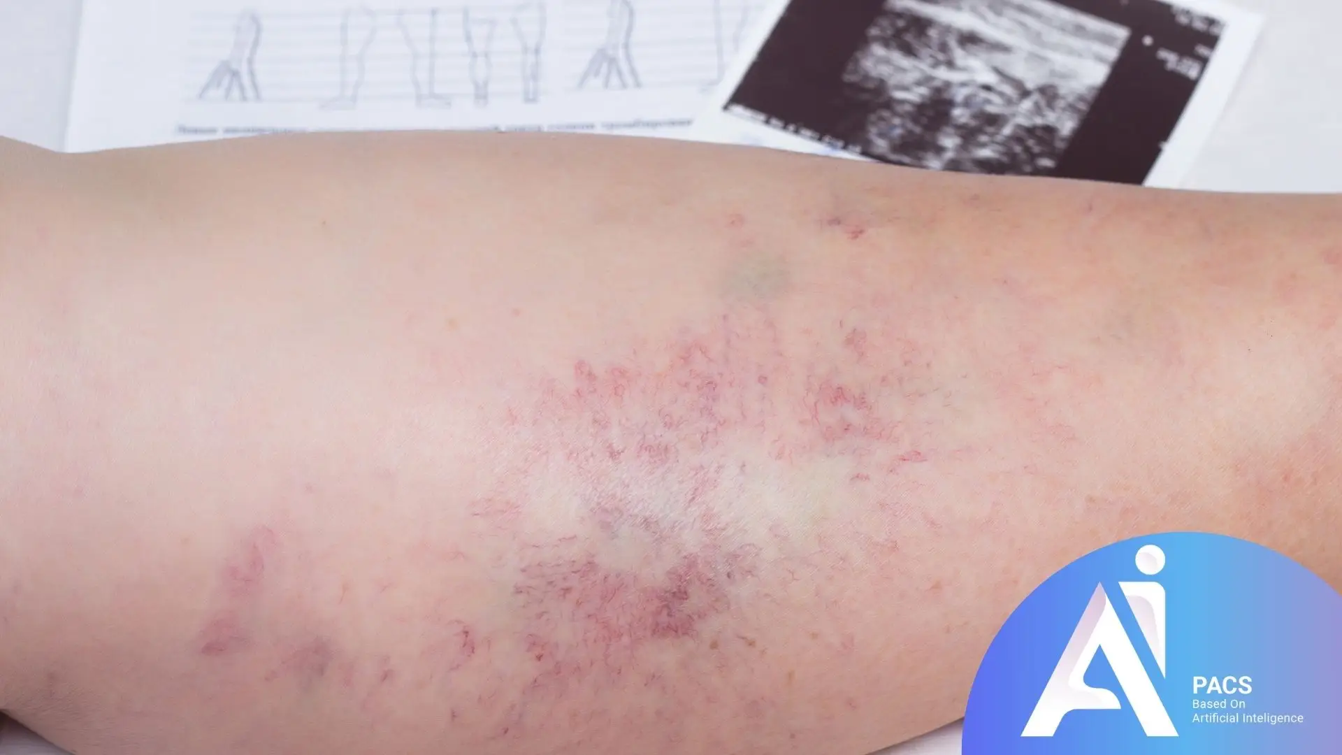
Common Symptoms of DVT
DVT can present with a variety of symptoms, which may vary in intensity depending on the severity of the clot. Common signs include:
- Swelling: Persistent swelling, often in one leg, is a hallmark symptom of DVT. The swelling may extend from the calf to the ankle or thigh.
- Pain or Tenderness: A dull ache or sharp pain, especially when walking or standing, can indicate DVT. The pain often begins in the calf.
- Redness or Discoloration: The skin around the affected area may appear red or discolored, often accompanied by warmth to the touch.
- Visible Veins: The veins near the skin’s skins may appear more prominent or bulging.
- Fatigue or Heaviness: Some individuals experience a sensation of heaviness or fatigue in the affected leg.
These symptoms may develop gradually or suddenly, depending on the size and location of the clot.
When to Seek Medical Attention
It is crucial to consult a healthcare provider immediately if you experience symptoms that may indicate DVT. Seek urgent medical attention if:
- Swelling or pain in the leg worsens rapidly.
- There is sudden, unexplained redness or warmth in the leg.
- You notice shortness of breath, chest pain, or rapid heartbeat—these could indicate a pulmonary embolism, a potentially life-threatening condition caused by a dislodged clot.
- You have a history of DVT or are at high risk due to factors such as recent surgery, immobility, or certain medical conditions.
Early evaluation with an ultrasound can confirm or rule out DVT, enabling timely treatment and reducing the risk of complications. Never ignore symptoms, as DVT can escalate quickly and lead to severe outcomes.
Ultrasound Techniques for Diagnosing DVT
Ultrasound is the most effective and widely used method for diagnosing Deep Vein Thrombosis (DVT). It employs different techniques to provide detailed visuals of the veins and assess blood flow, helping healthcare professionals detect blood clots accurately. Among these techniques, compression ultrasound and Doppler ultrasound are the most prominent, each serving a specific role in DVT diagnosis. Additionally, the ability of ultrasound to provide real-time imaging further enhances its diagnostic capabilities.

Compression Ultrasound (CUS)
The Gold Standard: Compression is the primary and most reliable technique for diagnosing DVT on ultrasound. This method involves applying gentle pressure with a transducer over the veins to assess their compressibility. Healthy veins collapse under pressure, but veins with clots remain rigid and non-compressible, indicating the presence of a thrombus. CUS is particularly effective for detecting clots in superficial and proximal deep veins, such as the thigh. Its simplicity, accuracy, and non-invasive nature make it the gold standard for DVT diagnosis.
Doppler Ultrasound
Doppler ultrasound complements compression ultrasound by focusing on blood flow dynamics within the veins. It uses sound waves to evaluate the speed and direction of blood flow, making it easier to identify blockages caused by clots. This technique is beneficial for detecting abnormalities in veins that are more profound in the body, where compression methods may be less effective. Doppler ultrasound can also assess the severity of DVT by identifying areas with reduced or absent blood flow, aiding in treatment planning.
Advantages of Real-Time Imaging
Ultrasound is a valuable tool for diagnosing deep vein thrombosis (DVT) due to its real-time imaging capabilities. This allows healthcare professionals to instantly observe blood flow, assess vein compressibility, and identify clots during the procedure. Real-time imaging also enables dynamic evaluations, such as testing for spontaneous blood flow, which provides additional insights into vascular health and supports immediate clinical decision-making.
Combined with real-time imaging, techniques like compression and Doppler ultrasound make diagnosing DVT fast and effective.
Interpreting Ultrasound Results for DVT
Ultrasound results confirm or rule out Deep Vein Thrombosis (DVT). Through detailed imaging, doctors can identify abnormalities in the veins, assess blood flow, and evaluate the extent of clot formation. Proper interpretation of these results is essential for determining the severity of the condition and planning appropriate treatment.
What Doctors Look for in Ultrasound Scans
When interpreting ultrasound scans for DVT, doctors focus on specific indicators that signal the presence of a blood clot. These include:
- Non-Compressible Veins: A vein that fails to collapse under pressure is a primary sign of DVT.
- Echogenic Thrombus: On the scan, the clot appears as a solid, echogenic (bright) structure within the vein.
- Distended Vein: A vein with a clot often appears swollen or enlarged compared to adjacent veins.
- Absence or Reduction of Blood Flow: Doppler ultrasound reveals areas where blood flow is restricted or completely absent.
- Venous Reflux or Spontaneous Flow Loss: Abnormalities in blood flow patterns, such as reverse or lack of spontaneous flow, further support the diagnosis.
Differentiating Between Acute and Chronic Thrombosis
A critical aspect of interpreting ultrasound results is distinguishing between acute (recent) and chronic (long-standing) thrombosis. This differentiation is crucial for guiding treatment and understanding the patient:
Acute Thrombosis:
- The vein is often distended and tender.
- The thrombus appears hypoechoic (less bright) on the ultrasound.
- The clot is usually non-adherent to the vein wall and may be mobile.
- Blood flow is significantly obstructed, with reduced or absent Doppler signals.
Chronic Thrombosis:
- The vein may appear smaller or have wall thickening due to scarring.
- Thrombus is echogenic (brighter) and firmly adherent to the vein wall.
- Collateral circulation (new smaller veins bypassing the clot) may be visible.
- Blood flow may persist but is altered due to vein damage.
Preventing DVT and Maintaining Vascular Health
Preventing Deep Vein Thrombosis (DVT) is crucial for maintaining vascular health and reducing the risk of serious complications like pulmonary embolism or chronic venous insufficiency. While some risk factors for DVT are unavoidable, such as genetics or medical conditions, there are effective strategies to minimize the likelihood of clot formation. Lifestyle changes and proactive healthcare practices are essential for prevention, particularly for high-risk patients.
Lifestyle Modifications to Lower DVT Risk
Adopting healthier lifestyle habits can significantly lower the risk of developing DVT. Key modifications include:
- Staying Active: Regular exercise improves circulation and prevents blood from pooling in the veins. Simple activities like walking, stretching, or swimming are beneficial.
- Avoiding Prolonged Sitting or Standing: For those who spend extended periods in a seated position, such as during long flights or desk jobs, taking breaks to move around or perform leg stretches helps maintain proper blood flow.
- Maintaining a Healthy Weight: Obesity increases pressure on the veins, so weight management can reduce strain and improve vascular health.
- Staying Hydrated: Adequate hydration prevents blood from becoming overly thick, reducing the risk of clot formation.
- Quitting Smoking: Smoking damages blood vessels and increases the likelihood of clots. Quitting smoking promotes overall vascular health.
- Wearing Compression stockings improves blood flow in the legs, particularly for individuals with varicose veins or a history of DVT.
Importance of Regular Check-Ups for High-Risk Individuals
For people at higher risk of DVT, regular medical check-ups are essential to monitor vascular health and address potential problems early. High-risk groups include those with a family history of DVT, previous blood clots, chronic illnesses, or prolonged immobility due to surgery or other conditions.
- Routine Screenings: Ultrasounds and other diagnostic tools can help detect early signs of DVT before symptoms appear.
- Medical Advice: Doctors can recommend personalized preventive measures, including medications like low-dose anticoagulants for high-risk patients.
- Monitoring Existing Conditions: Managing underlying issues, such as diabetes, heart disease, or inflammatory disorders, can prevent complications that contribute to clot formation.
Conclusion
Deep Vein Thrombosis (DVT) is a serious condition that needs prompt diagnosis and management to prevent life-threatening complications. Ultrasound is the gold standard for detecting DVT, providing accurate and non-invasive imaging to guide treatment.
To reduce the risk of DVT, especially in high-risk individuals, it’s important to maintain a healthy lifestyle, stay active, and attend regular medical check-ups. Combining advanced diagnostic tools with proactive health practices helps patients manage their vascular health effectively.