

Applications of Fluoroscopy
Fluoroscopy is an advanced imaging technique that allows physicians to obtain real-time, live views of internal organs and tissues. This method utilizes X-rays that pass through the body and project moving images onto a monitor. First introduced in the late 19th century, fluoroscopy has since undergone significant advancements, making it an indispensable tool in modern medicine. It is widely used in diagnostic and interventional procedures, offering valuable insights that assist in patient care.
Fluoroscopy is widely used in various medical fields, including:
Gastrointestinal Diagnosis and Treatment
It helps evaluate the function of the digestive system and detect conditions such as obstructions, ulcers, and tumors. It is commonly used in procedures like barium swallow, upper gastrointestinal series, and small bowel follow-through to assess digestive tract disorders.
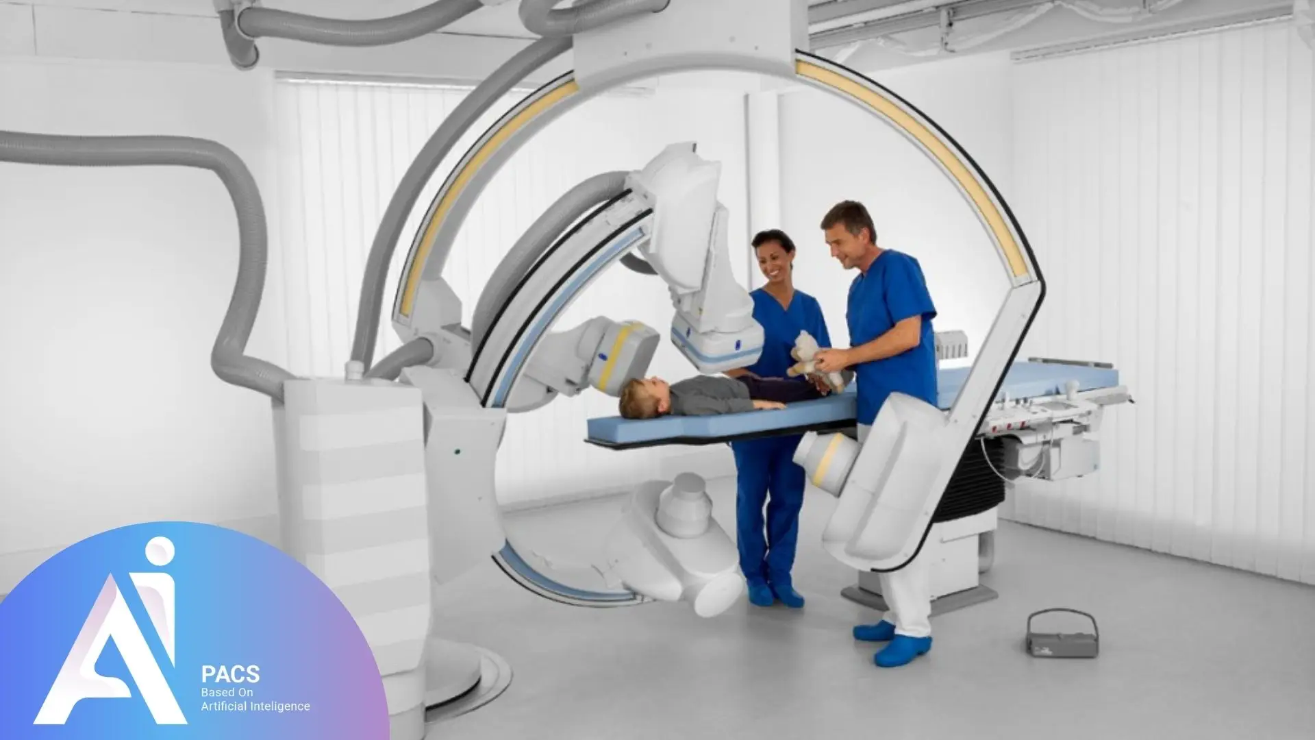
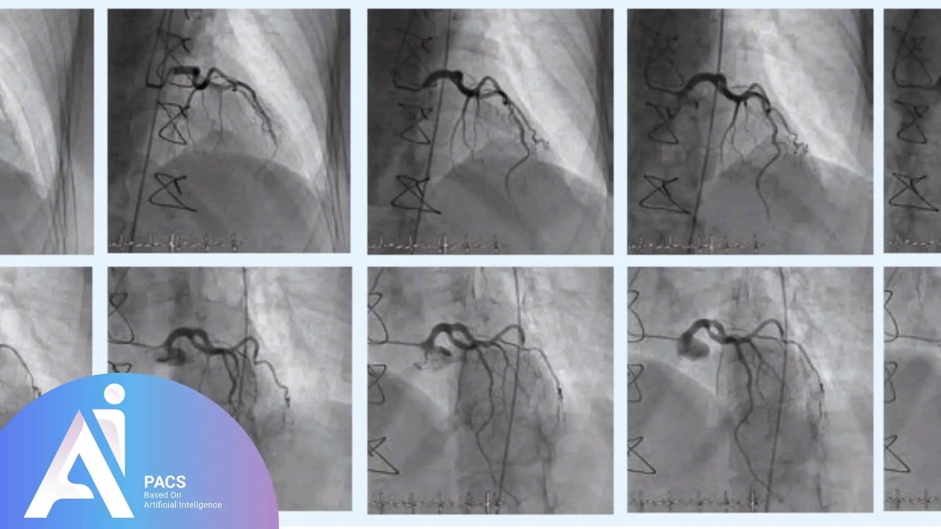
Cardiovascular and Vascular Examinations
This technique is essential in angiography, allowing doctors to visualize blood vessels and diagnose cardiovascular diseases. It plays a key role in catheter placements, stent insertions, and embolization procedures.
Surgical Guidance
In complex surgeries, fluoroscopy assists surgeons in precisely navigating surgical instruments. It is particularly useful in orthopedic procedures, spinal surgeries, and pain management injections.
Joint and Bone Assessments
It is used to examine joint and bone disorders, including fractures and dislocations. It also aids in diagnosing arthritis, ligament injuries, and guiding joint injections.
Wondering if fluoroscopy is right for your condition? 📁 Upload your imaging report now for a detailed second opinion from expert radiologists on AI-PACS.
Advantages of Fluoroscopy
Fluoroscopy offers several benefits, including:
✔ Real-Time Visualization of Internal Organs: Provides live imaging of internal structures, aiding in accurate diagnosis.
✔ Fast and Precise Diagnosis: Enables physicians to quickly and accurately identify internal abnormalities, leading to faster treatment plans.
✔ Reduced Need for Invasive Surgery: Many conditions can be diagnosed and treated without the need for open surgery, minimizing risks and recovery time.
✔ Enhanced Precision in Treatments: Allows for more targeted and effective medical procedures, ensuring better patient outcomes.
✔ Versatility: Fluoroscopy is adaptable across various medical specialties, from cardiology and gastroenterology to orthopedics and pain management.
Challenges and Limitations
Despite its advantages, fluoroscopy comes with certain challenges and limitations:
Radiation Exposure: The use of X-rays can be potentially harmful, making it crucial to regulate exposure levels and implement protective measures such as lead shielding and dose optimization techniques.
Requirement for Specialized Skills: Performing fluoroscopy demands expertise to ensure accurate and safe results. Proper training and certification are necessary for radiologic technologists and physicians to operate the equipment effectively.
High Costs: Fluoroscopy equipment is expensive, and maintenance and operation costs can also be significant. The need for advanced imaging technology and skilled personnel further adds to the overall expense.
Fluoroscopy Procedure: Step by Step
As mentioned earlier, fluoroscopy is a medical imaging process that involves the use of X-rays and a fluoroscopic screen. Below are the key steps involved in the procedure:
1. Patient Preparation
Patients are typically required to remove their clothing and wear a medical gown. Depending on the type of fluoroscopy, they may need to consume contrast agents like barium (for gastrointestinal studies) or receive an injected contrast medium (for vascular and cardiac examinations). Patients with allergies or kidney issues may require special precautions before receiving contrast agents.
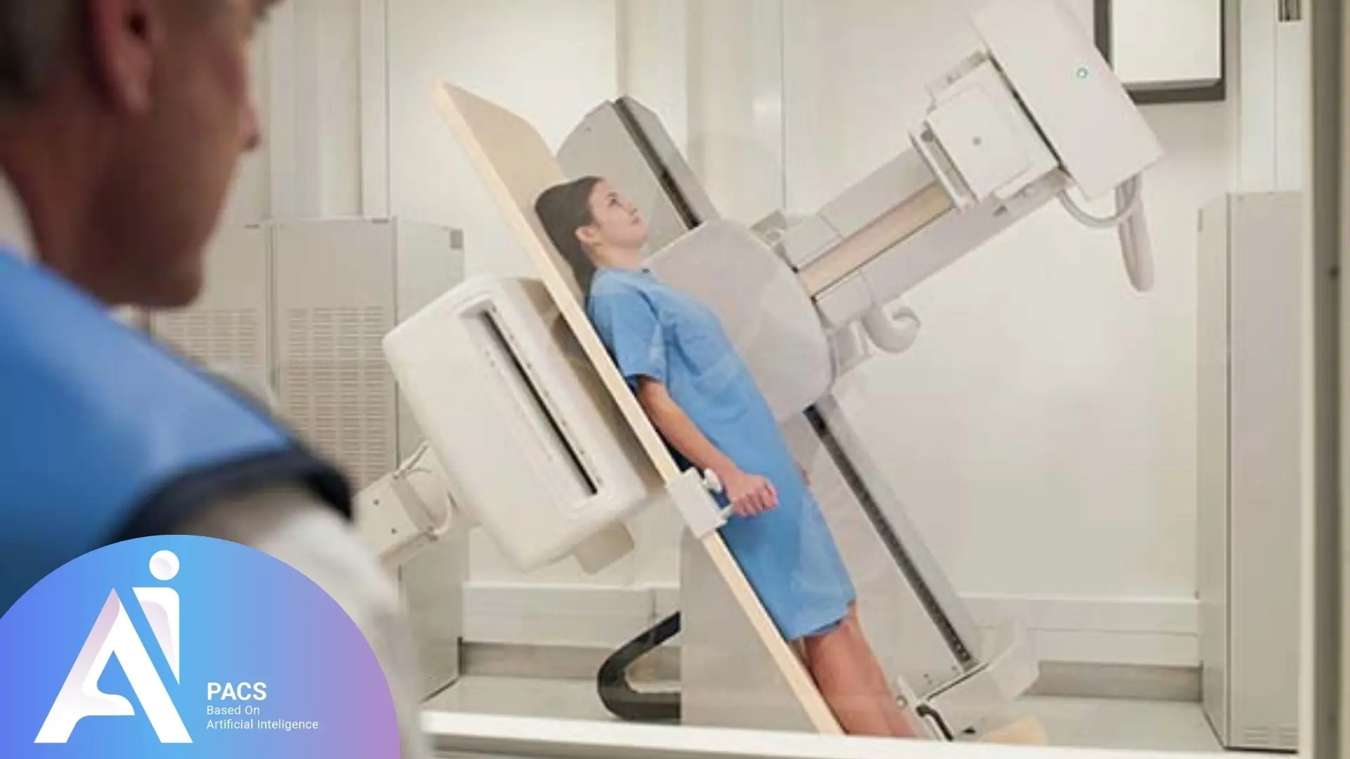
2. Patient Positioning
The patient is positioned on a fluoroscopy table. The exact position depends on the type of imaging being performed. The doctor may instruct the patient to maintain specific postures or change positions during the procedure to optimize image clarity.
3. Equipment Setup
Fluoroscopy equipment consists of an X-ray source and a fluoroscopic screen connected to a display system. The physician or radiologic technologist adjusts the equipment to achieve the best image angle and clarity while ensuring minimal radiation exposure.
4. Imaging Process
X-ray beams pass through the patient’s body and reach the fluoroscopic screen, transmitting live images to the display monitor. This enables real-time visualization of internal structures, allowing immediate assessment of function and abnormalities.
5. Image Observation and Recording
The physician observes the live images, recording videos or capturing still images when necessary. The patient may be asked to hold their breath or perform specific movements to obtain clearer images. The recorded images may be used for further analysis, treatment planning, or comparison in follow-up evaluations.
6. Completion of the Procedure
Once the imaging is complete, the patient can change back into their clothes and leave. If contrast materials were used, the doctor may provide post-procedure instructions, such as drinking plenty of water to help eliminate the contrast agent from the body. In some cases, follow-up imaging or blood tests may be recommended to monitor the patient’s response.
Fluoroscopy enables physicians to accurately detect internal conditions in real-time and determine the most appropriate treatment plans, improving patient management and care quality.
Fluoroscopy is one of the most powerful tools in medical imaging, but interpreting its results correctly is crucial. 👉 Get a professional second opinion on your fluoroscopy images through AI-PACS.
Final Thoughts
Although fluoroscopy is a non-invasive and relatively simple procedure, precautions must be taken to minimize unnecessary radiation exposure. Healthcare professionals ensure that the process is conducted safely while maintaining patient comfort. Patients should be informed about potential risks and benefits before undergoing the procedure.
Overall, fluoroscopy plays a crucial role in medical imaging, significantly enhancing the accuracy of diagnosis and treatment. By providing real-time visualization of internal structures, it enables doctors to make more precise clinical decisions and improve patient outcomes. Its application in multiple specialties continues to expand, reinforcing its importance in modern medicine.
Read MRI vs. CT Scan: Key Differences and Best Uses for Diagnosis to understand which imaging method suits your medical needs.
Reference:
my.clevelandclinic
hopkinsmedicine
howradiologyworks
cdc.gov