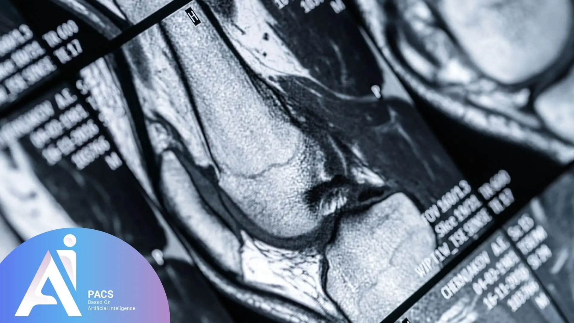
Understand How to Read Your Knee MRI Report
A knee MRI is ordered by a physician for various reasons. Here are some key purposes for performing this imaging study:
Soft Tissue Injury Diagnosis: MRI accurately detects injuries to ligaments, tendons, menisci, and cartilage. Examples include meniscal tears, anterior cruciate ligament (ACL) and posterior cruciate ligament (PCL) tears, and tendon injuries.
Evaluation of Inflammation and Infections: MRI can identify signs of inflammation, infections, or other inflammatory changes in the knee joint, such as bursitis, arthritis, and synovitis.
Bone Abnormalities and Structural Changes: This imaging method helps detect bone fractures, cartilage damage, and degenerative changes such as osteoarthritis.
Detection of Tumors and Cysts: MRI can reveal abnormal masses, tumors, cysts, and other irregularities within the knee joint.
Pre- and Post-Surgical Assessment: It is used to evaluate the knee before surgery and monitor post-surgical recovery and outcomes.
Vascular Abnormalities: MRI can identify vascular conditions such as thrombosis and vascular malformations.
Key Medical Terminologies in Knee MRI
Understanding these terms is essential for patients as it allows them to better comprehend their MRI reports, communicate effectively with healthcare providers, and make informed decisions about their treatment options.
Knee MRI reports contain numerous specialized medical terms that help in the precise diagnosis of knee conditions. Below are some common terms and their meanings:
- ACL (Anterior Cruciate Ligament): A crucial ligament in the knee that provides stability to the joint.
- PCL (Posterior Cruciate Ligament): A ligament located at the back of the knee, contributing to its stability.
- Meniscus: Two crescent-shaped cartilages in the knee joint that help absorb shock and provide stability.
- MCL (Medial Collateral Ligament): A ligament on the inner side of the knee that prevents excessive lateral movement.
- LCL (Lateral Collateral Ligament): A ligament on the outer side of the knee that supports lateral stability.
- Chondromalacia: Softening or degeneration of knee cartilage, usually beneath the patella (kneecap).
- Osteoarthritis: A degenerative joint disease that leads to cartilage breakdown and bone damage in the knee.
- Effusion: Excess fluid accumulation within the knee joint, typically due to inflammation or injury.
- Baker’s Cyst: A fluid-filled sac behind the knee, often caused by joint inflammation or injury.
- Bone Marrow Edema: Swelling or inflammation within the bone marrow, usually indicating injury or inflammation.
- Patellar Tendonitis: Inflammation of the patellar tendon, commonly associated with overuse or strain.
- Subchondral Cyst: A fluid-filled sac beneath the cartilage, often resulting from osteoarthritis.
- Ligamentous Tear: A partial or complete tear of one of the knee ligaments.
- Meniscal Tear: A tear in the meniscus, which can cause pain, swelling, and restricted knee movement.
- Synovitis: Inflammation of the synovial membrane that lines the knee joint, often seen in arthritis or joint injuries.
Post-MRI Actions
After completing a knee MRI, it is important to follow up with your physician or specialist to discuss the results. If abnormalities are detected, additional tests such as X-rays or blood tests may be recommended to provide a comprehensive diagnosis. In cases of injury or degenerative conditions, your doctor may suggest physical therapy, medication, or surgical options based on the MRI findings. Additionally, adopting lifestyle changes such as maintaining a healthy weight, engaging in low-impact exercises, and following a proper rehabilitation plan can aid in recovery and reduce future complications. Ensuring proper follow-up care can help manage symptoms and prevent further complications.
Final Thoughts
Early diagnosis through MRI plays a crucial role in preventing long-term joint damage and facilitating timely interventions. Identifying knee issues at an early stage allows for effective treatment planning, potentially avoiding invasive procedures and promoting faster recovery.
The interpretation of a knee MRI is performed by a radiologist, who carefully examines the images to identify any injuries, inflammation, or structural abnormalities. This evaluation plays a critical role in diagnosing conditions and determining an appropriate treatment plan. MRI results help physicians decide whether a patient requires surgery, physical therapy, or other non-invasive treatments. Given its high accuracy and detailed imaging capabilities, a knee MRI significantly contributes to improving patient outcomes and preventing severe complications. Therefore, a thorough and precise interpretation of MRI findings is essential for successful diagnosis and treatment.
💡 Tip: Keep a copy of your MRI report and images. These are valuable for second opinions or future comparisons.
References: