How to Interpret a Shoulder MRI Report
Shoulder MRI is a crucial diagnostic tool used to evaluate a range of musculoskeletal conditions, including rotator cuff tears, labral injuries, and joint degeneration. It provides high-resolution imaging to aid in the detection of soft tissue injuries, joint disorders, and bone abnormalities. For example, if someone experiences persistent shoulder pain after a fall, an MRI can help determine if they have a torn rotator cuff or damage to the labrum—conditions that might not show up clearly on an X-ray. By offering detailed images of internal structures, MRI allows healthcare professionals to diagnose conditions that might not be easily identified through other imaging methods, such as X-rays or ultrasound. This article explores the key applications, advantages, and terminology associated with shoulder MRI interpretation.
Applications of Shoulder MRI
1. Diagnosis of Soft Tissue Injuries
Shoulder MRI is essential for identifying injuries to soft tissues like tendons, ligaments, and muscles. For example, a tennis player who experiences sharp pain while serving may have a tear in the rotator cuff tendon. An MRI scan can reveal the extent of the damage, whether it’s a small partial tear or a full‑thickness tear, helping doctors determine whether conservative treatment like physical therapy will work or if surgery is necessary.
If your MRI shows a possible tendon tear and you want clarity on whether surgery is needed or not, submit your MRI report to AI-PACS for an expert second opinion.
2. Assessment of Joint Disorders
MRI is also crucial for diagnosing joint disorders such as arthritis, inflammation, and instability in the shoulder. For instance, a person in their 60s who experiences stiffness and difficulty moving the shoulder may have osteoarthritis, where the cartilage in the shoulder joint is wearing down. MRI can detect the degree of cartilage loss and show any bone spurs, giving the doctor important information for recommending treatments like injections or possibly joint replacement.
3. Evaluation of Bone Injuries
When it comes to evaluating bone injuries, shoulder MRI excels in detecting fractures and bone lesions that might not be visible on an X‑ray. To make it clear, consider an athlete who falls during a game and hurts their shoulder may have a fracture in the clavicle (collarbone). While an X‑ray might not show the full extent of the injury, an MRI can provide detailed images, revealing any fractures and ensuring that doctors can assess any damage to nearby soft tissues, such as tendons or ligaments.
4. Tumor Detection
MRI technique is also highly effective for detecting tumors or other abnormal growths in the shoulder area. If a person notices unexplained swelling or pain, an MRI scan can identify whether the issue is a benign tumor, like a lipoma (a fatty tumor), or something more serious, such as bone cancer. Early detection can make a significant difference in treatment outcomes, ensuring that any necessary interventions, like surgery, are done promptly.
5. Post-Surgical Assessment
After shoulder surgery, MRI is an excellent tool for assessing the healing process. Whether someone has undergone rotator cuff repair or labral tear surgery, the scan can verify if the repair is successful, and if there are any complications such as scar tissue formation or misaligned implants (e.g., a person who had surgery to repair a torn rotator cuff can undergo an MRI to ensure the tendon is healing properly and that no new tears have developed).
6. Evaluation of Chronic Shoulder Pain
Chronic shoulder pain can be difficult to diagnose, especially when other treatments like physical therapy or medication haven’t helped. MRI can identify underlying causes that may not be obvious during a physical exam. To put it into perspective, someone suffering from long‑term shoulder pain might be diagnosed with frozen shoulder (adhesive capsulitis) or a labral tear, both of which can be clearly seen on an MRI. Understanding the exact cause of the pain allows doctors to create a more effective treatment plan.
7. Assessment of Nerve Compression
Nerve compression in the shoulder can cause symptoms like numbness, tingling, or weakness in the arm. MRI is a powerful tool for detecting nerve impingements or other conditions that might be pinching nerves. Take, for instance, someone experiencing tingling or weakness in the arm when lifting heavy objects may have a pinched nerve in the neck or shoulder area. MRI can show where the nerve is being compressed, helping doctors determine if it’s due to a herniated disc, shoulder issue, or other underlying conditions.
Shoulder Anatomy and Variants on MRI
Supporting Structures of the Glenohumeral Joint
The glenohumeral joint is supported by various structures that help stabilize and facilitate movement. Here’s a breakdown of those supporting structures:
Superiorly:
- Coracoacromial Arch and Coracoacromial Ligament: These form a protective arch over the shoulder joint, helping prevent upward displacement of the humeral head (This is like a roof that shields the top part of the shoulder joint).
- Long Head of the Biceps Tendon: The tendon that attaches the biceps muscle to the shoulder, helping in both stabilization and movement (Think of this tendon as a rope that connects the biceps muscle to the shoulder joint, aiding in arm movement and stability).
- Tendon of the Supraspinatus Muscle: A key muscle tendon in the rotator cuff, it helps lift the arm (This tendon allows you to raise your arm out to the side, such as when reaching for something on a high shelf).
Anteriorly:
- Anterior Labrum: A piece of cartilage that forms a cup around the ball of the shoulder joint, stabilizing it (This is like a cushion around the ball of the shoulder, making sure it stays in place).
- Glenohumeral Ligaments – SGHL, MGHL, IGHL (Anterior Band): These ligaments connect the shoulder bones and keep the joint stable during arm movements (These are strong bands that help keep the shoulder joint together, like a seatbelt for the shoulder).
- Subscapularis Tendon: Part of the rotator cuff, this tendon helps rotate the arm inward (This tendon lets you rotate your arm, such as when turning a doorknob).
Posteriorly:
- Posterior Labrum: Another piece of cartilage, but located in the back of the shoulder, helping stabilize the joint (Like the cushion in the front, but this one is at the back of the shoulder to keep everything in place).
- Posterior Band of the IGHL: A ligament that contributes to the shoulder’s stability by preventing dislocation at the back of the joint (This band helps prevent your shoulder from moving too far backward, keeping it secure).
- Infraspinatus and Teres Minor Tendon: Tendons of two muscles in the rotator cuff that allow the arm to rotate outward (These tendons help you rotate your arm outward, like when reaching behind you or throwing a ball).
Key Points and What’s Being Analyzed
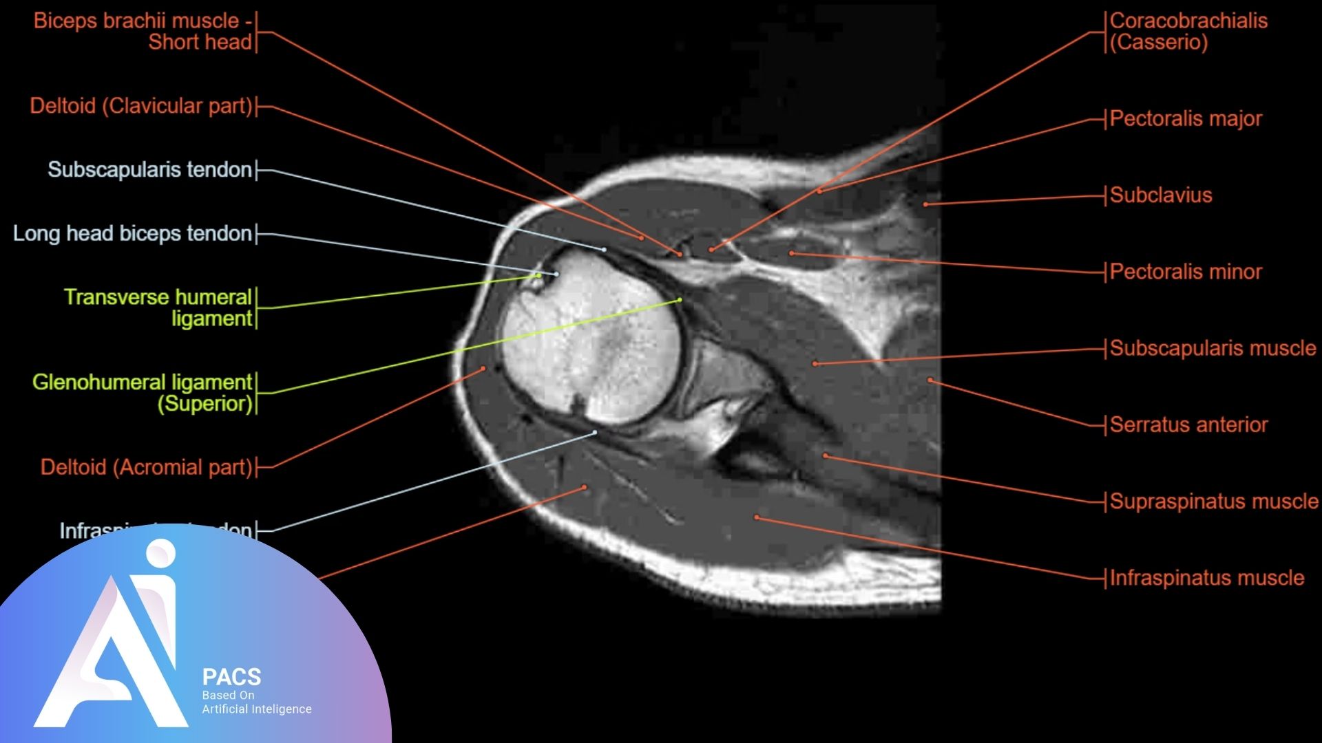
Axial Anatomy:
- Os Acromiale: This refers to a condition where part of the acromion bone in the shoulder remains separated. The purpose of examining this is to see if there’s any abnormality in the bone structure that could affect shoulder movement or cause pain.
- Supraspinatus Tendon Alignment: The supraspinatus tendon is part of the rotator cuff, and it’s analyzed to make sure it’s properly aligned with the muscle. If it’s not, it could be a sign of injury or other shoulder issues.
- Biceps Tendon Position: The biceps tendon attaches at the top of the shoulder. The purpose of reviewing this is to ensure it’s in the correct position, as a misalignment could be related to shoulder instability or pain.
- Superior Labrum and Glenohumeral Ligament: These are structures that help keep the shoulder joint stable. The evaluation focuses on identifying any damage or tears that could affect shoulder movement, such as SLAP lesions (tears in the labrum) or a sublabral foramen (a small hole in the cartilage).
- Hill-Sachs Lesion: This is a type of injury that can occur when the shoulder dislocates. The diagnostic imaging helps identify if there is any damage to the humeral head (the ball of the shoulder) that could cause ongoing problems.
- Subscapularis Tendon: This tendon helps keep the biceps tendon in its proper place. Reviewing this tendon ensures it’s intact and functioning correctly, which is essential for shoulder stability.
- Glenohumeral Ligaments (GHL): These ligaments help keep the shoulder joint stable. Assessing them helps identify any damage or abnormalities that could cause shoulder instability.
- Normal Contour of the Humeral Head: The shape of the humeral head is analyzed to make sure it hasn’t been deformed, which could happen due to shoulder dislocations or other injuries.
- Bankart Lesions: This is a tear in the cartilage that helps hold the shoulder joint in place. The MRI imaging helps detect any damage that could lead to recurring shoulder dislocations.
- Inferior GHL Fibers: These fibers help hold the shoulder joint in place. Assessing them helps identify any potential issues that could affect the shoulder’s stability.
Supraspinatus Tendon:
- The supraspinatus tendon is a key part of the rotator cuff and helps you lift your arm. It’s commonly injured, so the MRI results focus on identifying tears or inflammation (tendinopathy). This tendon is most clearly seen on special views called coronal oblique and ABER-series. These images are used to focus specifically on the tendon’s condition.
Coronal Anatomy:
- Coracoclavicular Ligament and Biceps Tendon: These structures are analyzed to ensure they are properly connected and functioning. Any issues here could affect shoulder stability.
- Coracoacromial Ligament: This ligament helps stabilize the shoulder. Reviewing it is important to identify any thickening or changes that could lead to shoulder impingement (pain caused by compression of the tendons).
- Suprascapular Nerve and Vessels: These nerves and blood vessels are essential for shoulder function. The assessment focuses on ensuring there is no nerve damage or poor blood flow affecting the shoulder.
- Supraspinatus Impingement: The diagnostic images look for signs that the supraspinatus tendon is being pinched or irritated by the bone structures, which can cause pain. This can happen due to bone spurs or a thickened ligament.
- Biceps-Labrum Complex: This area connects the biceps tendon to the labrum (cartilage in the shoulder). The examination focuses on detecting tears (SLAP tears) or other problems here that could lead to shoulder pain or instability.
- Subacromial Bursa and Supraspinatus Tears: The bursa is a fluid-filled sac that reduces friction in the shoulder. If there’s too much fluid in it, it could be a sign of inflammation or injury. The MRI evaluation also looks for tears in the supraspinatus tendon, a common injury.
- Rim-Rent Tears: These are tears in the tendon where it attaches to the bone. The diagnostic scan helps identify these tears to see if they are contributing to shoulder problems.
- IGHL and HAGL Lesions: These refer to tears in ligaments that help keep the shoulder joint stable. The MRI analysis looks for these lesions to ensure the shoulder joint is secure and not at risk of dislocation.
- Infraspinatus Tendon Tears: This tendon helps rotate the arm. Assessing it for tears is important because injuries here can limit arm movement.
- Hill-Sachs Lesion: A small Hill-Sachs lesion can appear after a shoulder dislocation. The MRI imaging focuses on detecting this to see if it’s affecting the shoulder’s function.
Sagittal Anatomy:
- Rotator Cuff Muscles: The rotator cuff muscles help stabilize the shoulder. The assessment focuses on checking these muscles for signs of atrophy (shrinking) that could affect shoulder movement.
- MGHL (Middle Glenohumeral Ligament): This ligament plays an important role in stabilizing the shoulder. The MRI evaluation looks at its condition to ensure the shoulder is stable.
- Labral Tears: At the 3-6 o’clock position, the MRI scan looks for any tears in the labrum, which could affect shoulder stability.
- Biceps Anchor: This is where the biceps tendon attaches to the shoulder. The MRI scan looks at this area to ensure it’s intact and functioning properly.
- Acromion Shape: The acromion is part of the shoulder blade. The imaging focuses on its shape to check for any abnormalities that could lead to shoulder impingement.
- Impingement by the AC Joint: The acromioclavicular (AC) joint is where the shoulder blade meets the collarbone. The MRI scan focuses on checking for signs of impingement (pain caused by the joint rubbing against other structures).
- Rotator Cuff Interval: The rotator cuff interval is an important area for shoulder stability. The MRI analysis checks this space for any problems that could affect the shoulder’s movement.
- Supraspinatus Tears: Finally, the MRI results focus on the supraspinatus tendon again for any tears or signs of injury, as it’s a critical tendon for shoulder movement.
By understanding these key areas and the purpose of checking them, you’ll have a clearer idea of what’s happening in your shoulder and whether there’s any injury or damage that needs attention.
Technical Terminology in Shoulder MRI Reports
Shoulder MRI images contain various technical terms used to describe anatomical structures, abnormalities, and injuries. Some of these terms include:
Terms Related to Abnormalities and Injuries
- Avascular Necrosis (Osteonecrosis): A condition where bone tissue dies due to a lack of blood supply, often detected in shoulder imaging, particularly when there is pain or limited movement.
- Bankart Lesion: A tear in the anterior labrum, frequently mentioned in shoulder scan findings related to shoulder dislocations or instability.
- Bursitis: Inflammation of the bursa, often identified in diagnostic imaging as an indication of rotator cuff issues or impingement.
- Capsulitis (Frozen Shoulder): Inflammatory condition of the shoulder capsule, often seen in MRI findings of patients with limited range of motion and pain.
- Cuff Tear Arthropathy: A type of shoulder arthritis that develops after a long-term rotator cuff tear, often identified in radiologic assessments.
- Dislocation: Shoulder dislocations are often mentioned in medical imaging when evaluating injuries or instability, particularly for labral tears or bone damage.
- Fracture: Breaks in the bones, especially the clavicle, humerus, or scapula, often evaluated through radiographic examinations to assess healing or related injuries.
- Impingement: Frequently mentioned in shoulder scans to describe the compression of rotator cuff tendons, especially in the subacromial space.
- Labrum Tears: Cartilage in the shoulder joint that provides stability; tears or degeneration of the labrum are commonly noted in shoulder imaging results.
- SLAP Tear: A specific type of tear in the superior labrum, often noted in diagnostic results when evaluating shoulder instability or pain.
- Subacromial Bursitis: Inflammation of the subacromial bursa, which is a fluid-filled sac that reduces friction between the acromion and the rotator cuff tendons.
- Tear: A common finding in shoulder imaging reports, often used to describe tendon or labral tears that impact shoulder function.
- Tendinosis/Tendonitis: Inflammation or degeneration of tendons, frequently mentioned in radiologic findings for conditions affecting the rotator cuff or biceps tendon.
- Winging (Scapula): Scapular winging may be noted in imaging studies when there is muscle imbalance or nerve damage affecting shoulder stability.
- Reverse Shoulder Prosthesis: When assessing post-surgical outcomes or planning for joint replacement, reverse shoulder prosthesis may be mentioned in medical imaging results.
- Shoulder Prosthesis / Arthroplasty: Imaging may reference shoulder replacements or artificial joints when evaluating the condition of the joint after surgery.
Final Thoughts
Early and accurate interpretation of MRI findings can significantly impact treatment outcomes, helping to determine whether conservative treatments are sufficient or if surgical intervention is necessary. Seeking professional evaluation ensures that the results are properly understood and that the best possible treatment plan is developed for each patient. A shoulder MRI requires specialized knowledge to accurately identify anatomical structures and abnormalities. High-resolution images enable physicians to diagnose soft tissue injuries, joint disorders, and bone lesions with precision. Understanding technical terms such as rotator cuff, labrum, and bursa is crucial in this process.
Additionally, patients should be aware that MRI findings should always be correlated with clinical symptoms and physical examination results to ensure an accurate diagnosis. A radiologist or orthopedic specialist will interpret the results and recommend the most appropriate treatment plan. Ultimately, a precise interpretation of shoulder MRI can aid in creating an effective treatment plan, facilitating faster patient recovery.
References: