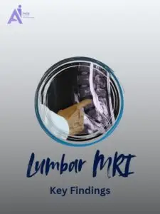
Understanding Lumbar Spine MRI Report: Key Insights and What to Expect
Have you received an MRI report for your lower back and found yourself overwhelmed by terms like bulging disc, herniation, or Modic changes? You’re not alone. MRI reports are often written for doctors, not patients, leaving many people confused and anxious.
MRI of the lumbar spine is commonly used to evaluate chronic or sudden back pain, leg numbness, or suspected nerve impingement. This guide will help you understand the key terms in your report so you can make more informed decisions about your health.
Real-Life Scenario: A Patient’s Journey
Sara, a 45-year-old office worker, had been suffering from lower back pain and tingling in her left leg for months. Her doctor ordered an MRI scan, which revealed findings like disc extrusion, foraminal narrowing, and Modic Type 2 changes. Unsure what this meant or whether she needed surgery, she contacted us for a clear explanation.
👉 Get Your Spine MRI Explained by an Expert
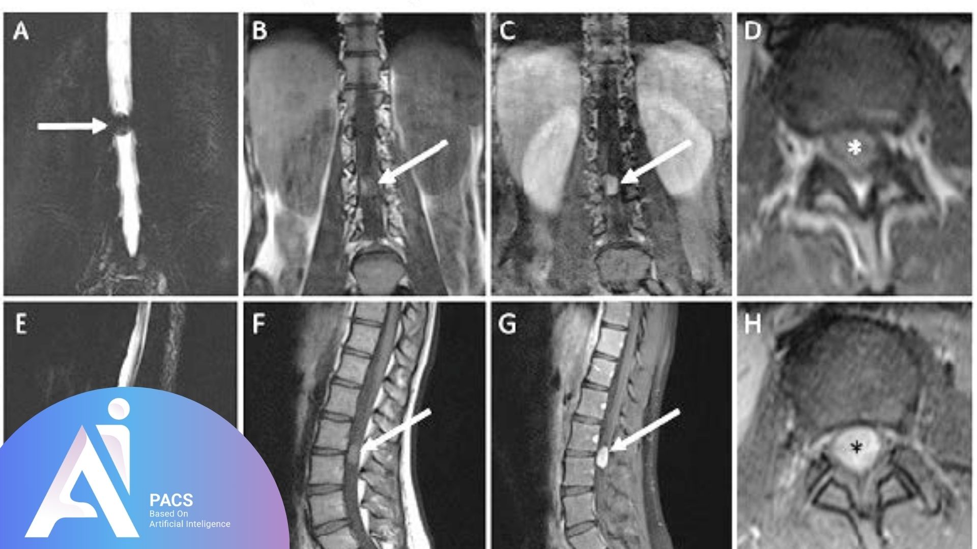
What Do These Medical Terms Mean?
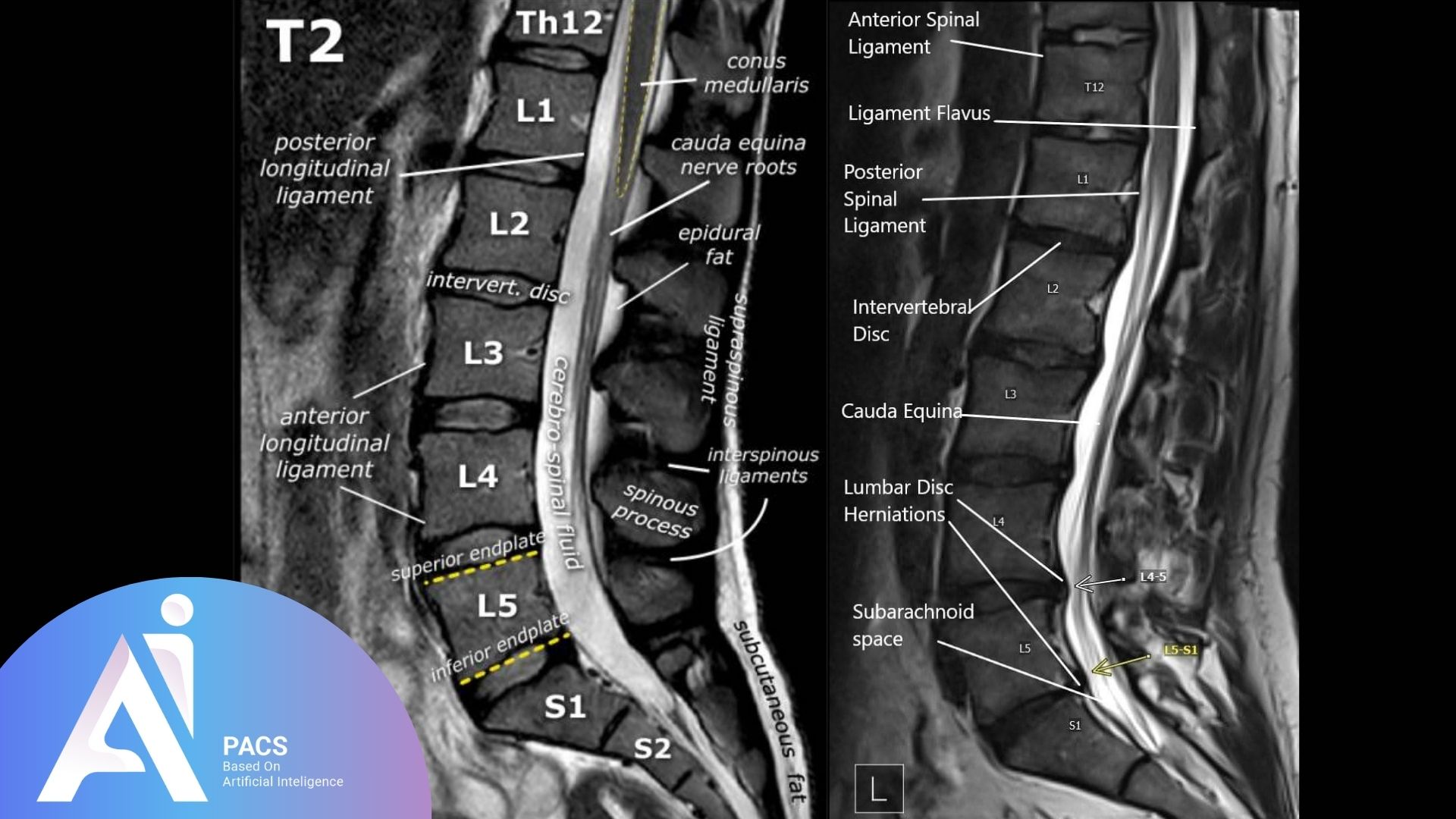
Intervertebral Disc
A cushion-like structure between vertebrae, made of a rigid outer ring (annulus fibrosus) and a soft gel-like center (nucleus pulposus).
Why It Matters: Discs absorb pressure and allow the spine to bend and move.
Disc Bulging
The disc extends outward beyond its normal space, but the outer ring remains intact.
Why It Matters: Common with aging, it may cause no symptoms unless it compresses nerves.
Treatment Needed? Usually not, unless symptoms are significant.
Disc Herniation
The inner gel leaks through a tear in the outer ring. Two main types:
Disc Protrusion
- The disc material bulges but remains partially contained.
- It may cause mild to moderate nerve symptoms.
Disc Extrusion
- The disc material entirely escapes the outer ring.
- Higher risk of severe symptoms; sometimes requires surgery.
Contained vs Uncontained Herniation
- Contained: The herniated material is still partially enclosed.
- Uncontained: The material has entered the spinal canal (often pressing the thecal sac or nerve roots).
Why It Matters: Uncontained extrusions are often more painful and less likely to improve without treatment.
Location of Herniation
- Central: Midline in the spinal canal.
- Paracentral: Just off center; most common.
- Foraminal: In the exit hole for spinal nerves.
- Lateral Recess: At the sides of the spinal canal near nerve roots.
Why It Matters: The location determines which nerve may be compressed.
Annular Fissure
A small tear in the disc’s outer ring without extrusion.
Symptoms: May cause localized pain or inflammation.
Treatment Needed? Often conservative, such as physical therapy.
Modic Changes
Degenerative changes in the vertebral endplates (bones adjacent to discs). Three stages:
- Type 1: Inflammatory (active pain).
- Type 2: Fatty replacement.
- Type 3: Bone hardening (sclerosis).
Why It Matters: Reflects chronic disc degeneration.
Facet Joints and Facet Arthropathy
Small joints in the back of the spine that control motion. Degeneration leads to arthritis (facet arthropathy).
Symptoms: Localized low back pain, especially with movement.
What Does This Report Say About My Condition?
MRI reports usually list findings as mild, moderate, or severe. Many people have abnormalities with no symptoms at all. Here’s how to interpret severity:
- Mild changes: Often age-related and not concerning.
- Moderate changes: These may cause symptoms but can often be treated conservatively.
- Severe changes may cause nerve compression and require further evaluation.
Important: The MRI must always be interpreted along with your symptoms. Not every disc problem means surgery.
Why You Should Get an Expert to Review Your Report
Interpreting your own report is risky. Here’s why:
- Some findings may sound serious, but are not.
- Others may be subtle yet important.
- Without expert input, you may misunderstand the urgency or overlook necessary care.
At AI-PACS, our team provides expert second opinions to help you understand your report clearly and guide your next steps.
Next Steps: Let’s Help You Understand Your Report
If you’ve received an MRI or CT scan report and aren’t sure what the findings mean, upload your report to our team of medical imaging experts.
📁 Submit your file here on AI-PACS online radiology report services
Deeper Dive Into Medical Imaging (clear, not overly technical)
1) Disc bulge vs herniation: the standard definitions
A bulge means the disc extends a little beyond its edges, but the outer ring remains intact. A herniation means nuclear material exits through a torn outer ring. Herniations are described as protrusion (wider base than tip) or extrusion (narrow base with material that can migrate). If the fragment detaches, it is called sequestration. These terms come from expert society consensus so surgeons, physiatrists, and radiologists all speak the same language [3].
2) Where symptoms come from: location matters
• Central canal: can press the sac of nerves and cause back pain or neurogenic claudication.
• Lateral recess: near the traversing nerve (for example, L5 at L4‑5).
• Foraminal/extraforaminal: at the nerve’s exit, often affecting the exiting root (for example, L4 at L4‑5).
Matching location to your symptoms and exam helps decide whether physical therapy, injection, or surgery is most likely to help [1][4].
3) Stenosis and severity words explained
Stenosis means narrowing. Reports may grade central canal, lateral recess, or foraminal stenosis as mild, moderate, or severe, often based on how much the nerves are crowded or displaced. Mild narrowing is common in people without pain. Severe stenosis with clear nerve compression and matching symptoms is more likely to need escalation of care [1][2][4].
4) Modic changes and endplates
Modic changes are MRI patterns in the bone just above and below a disc. Type 1 reflects active inflammation, Type 2 shows fatty change from chronic degeneration, and Type 3 indicates bony hardening. These changes can correlate with pain in some patients, but they are also seen in people without symptoms. They help explain chronicity and guide expectations, not automatic surgery [2].
5) Facet joints, ligamentum flavum, and spondylolisthesis
Facet arthropathy (arthritis of the small joints) and thickening of the ligamentum flavum can narrow the canal or foramina, adding to stenosis. A slip of one vertebra on another, called spondylolisthesis, may coexist. Facet effusions on MRI can hint at instability, which your clinician may evaluate with targeted exam and, sometimes, motion X‑rays [1][4].
Final Thoughts
Interpreting your lumbar spine MRI report can be overwhelming, but with the right guidance, it becomes much clearer. Not all findings indicate the need for immediate treatment, and many can be managed conservatively. Always consult a healthcare professional to ensure you understand your results and determine the best course of action. If you’re still unsure, our expert team is here to help you navigate your MRI findings and next steps for better spine health.
