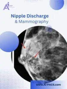

What Does a Mass on a Mammogram Mean?
A mass is an area with an outline that stands out from nearby breast tissue. Its appearance can raise or lower concern, but the word “mass” alone does not establish whether it is cancer. A finding on an image may not be something you can feel.
Mammography uses low-dose X-rays. Screening looks for changes before symptoms develop; diagnostic mammography investigates a symptom or a finding needing a closer look.

Cysts, Solid Masses and Other Findings
A fluid-filled cyst and a solid mass can look similar on a mammogram. Ultrasound can help tell them apart. A solid finding is not automatically malignant: fibroadenomas are one benign example. The radiologist uses the complete imaging assessment to recommend what happens next.
Which Features Does the Radiologist Assess?
The report may describe the mass’s location, size, shape, margins and density, associated calcifications, and changes from earlier imaging. No single adjective should be read as a cancer diagnosis. Sharing previous mammograms can help the radiologist assess whether a finding is new or has changed.

What Happens After a Mass Is Found?
You may be asked to return for diagnostic views or ultrasound. The next test depends on the finding and clinical question; MRI is used selectively, not automatically for every mass. When a finding is suspicious, a biopsy may be recommended so a pathologist can examine a sample. Imaging assessment and tissue diagnosis serve different purposes.
If you can feel a new or persistent lump, tell your clinician even if the imaging report is normal. Some symptoms need further evaluation despite reassuring images. Keep recommended appointments while arranging an independent review.
Read the BI-RADS Assessment and Recommendation
- 0: the assessment is incomplete; more imaging or comparison is needed.
- 1: no abnormality identified on this examination.
- 2: benign finding.
- 3: probably benign; short-interval follow-up is usually recommended.
- 4: suspicious finding; biopsy is generally recommended.
- 5: highly suggestive of malignancy; biopsy is needed to establish the diagnosis.
- 6: malignancy already confirmed by biopsy.
BI-RADS communicates the imaging assessment; it is not a cancer stage. Follow the recommendation attached to your result.
When an Independent Mammogram Review May Help
An independent review can reassess the images behind an unclear report or a recommendation you want explained. Read about mammogram second opinions for the workflow, or start a consultation request.
Ask your imaging center for the complete original DICOM study, including additional views and relevant prior images, plus the report if available. A PDF report or phone photograph alone is not enough for diagnostic image review. Ask the team to check file suitability and case scope before ordering. Another review may agree with the original assessment and does not replace your treating clinician or a recommended biopsy.
Still waiting for your first report? Contact the imaging center about its expected delivery. For an alternative reporting request, read online radiology reporting and confirm that the available files can be assessed. Do not postpone recommended clinical care while waiting.