


What is MR Spectroscopy?
We are all familiar with MRI images, which help doctors take a precise look inside our bodies. However, beyond these images, there is another technology capable of providing molecular-level information about body tissues.
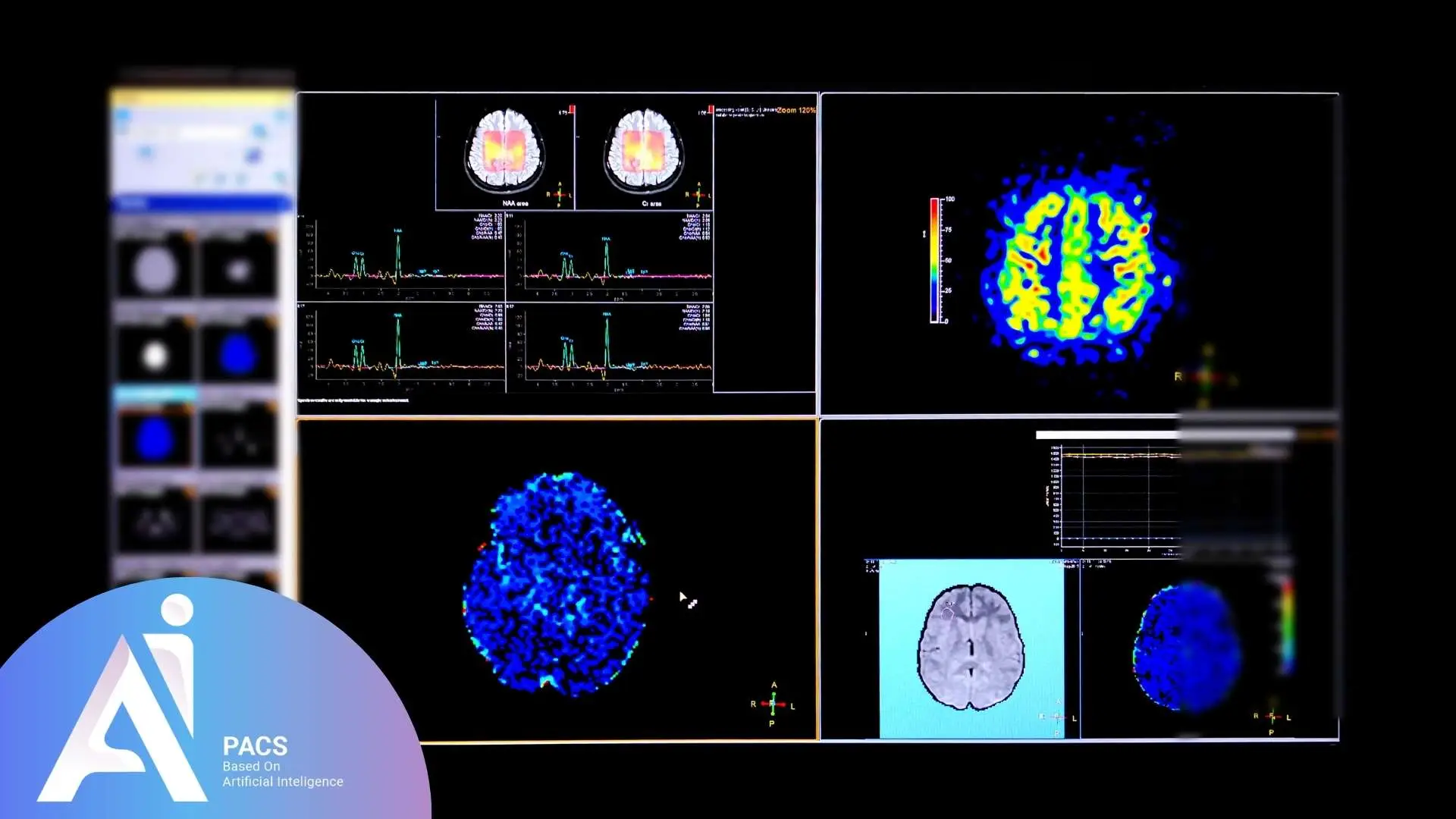
Magnetic Resonance Spectroscopy (MRS) is an advanced imaging technique derived from MRI that provides molecular-level insights into the chemical composition of cells and tissues, rather than focusing solely on structural imaging.
This non-invasive test, also known as non-invasive biopsy, identifies tissue composition, measures molecules within tissues, and compares the chemical composition of normal and abnormal (tumorous) tissues. MRS is used to examine pathological lesions in various organs, including the brain, prostate, and breast tissue, helping determine the type, size, and anatomical location of tumors. The primary objective of this technique is to analyze the chemical environment based on hydrogen atoms, enabling the identification of metabolites such as choline, N-acetyl aspartate (NAA), creatine, and lactate, which provide insights into various neurological and oncological conditions. MRS is particularly useful for assessing conditions such as seizures, tumors, strokes, multiple sclerosis (MS), Alzheimer’s disease, and other pathological conditions. It plays a crucial role in the early detection and diagnosis of cancers, infections, metabolic disorders, and many other diseases.
Looking for high-quality online radiology report services? Our AI-PACS team of specialists is here to provide you with precise evaluations.
Applications of MRS
MR Spectroscopy is primarily utilized to distinguish between malignant and benign tumors, detect metabolic changes in neurological disorders, and differentiate between tumor recurrence and radiation necrosis. It is also instrumental in detecting metabolic changes associated with neurological disorders such as epilepsy and neurodegenerative diseases.
Common Conditions Diagnosed with MR Spectroscopy
MR Spectroscopy is widely used to analyze metabolic changes in various conditions, especially in the brain. Common applications include:
- Brain tumors: Differentiating malignant tumors from benign growths and monitoring treatment response.
- Epilepsy: Identifying seizure foci by detecting abnormal metabolic patterns.
- Multiple sclerosis (MS): Assessing disease activity by measuring changes in brain metabolites.
- Stroke: Evaluating tissue damage and recovery by detecting biochemical changes.
- Neurodegenerative diseases, Such as Alzheimer’s, can be revealed by altered brain chemistry before structural changes appear.
This technology enables doctors to understand the underlying biochemical environment, resulting in more accurate diagnoses and personalized treatments.
Why is MR Spectroscopy Prescribed?
MRS is commonly recommended for:
- Diagnosing and monitoring specific diseases
- Determining the type and anatomical location of tumors
- Measuring metabolism and detecting chemical changes in tissues
- Comparing the chemical composition of normal brain tissue with abnormal tumor tissue
- Assessing metabolic changes in conditions like multiple sclerosis, Alzheimer’s, and epilepsy
Advantages of MRS
The benefits of Magnetic Resonance Spectroscopy (MRS) include:
- A non-invasive method that provides valuable information without requiring a tissue biopsy
- The ability to detect chemical changes in tissues before structural changes occur
- The capability to simultaneously examine multiple metabolic processes in different organs
- Used for distinguishing between malignant and benign tumors
- Helpful in evaluating the progression of neurodegenerative diseases

How to Prepare for MR Spectroscopy?
Generally, no special preparation is required. However, patients should discuss any medical conditions or implanted medical devices with their physician. This method is safe, but like any imaging technique, it should only be performed when the information obtained is essential for diagnosis or treatment.
To prepare for MRS, the following precautions may be necessary:
Avoiding Caffeinated Beverages: Patients may be advised to refrain from consuming caffeinated drinks before the test, as caffeine can affect brain metabolism.
Wearing Comfortable Clothing: Since the patient needs to remain in a lying position for approximately 30 minutes, wearing comfortable clothing is recommended.
Avoiding Jewelry and Metal Objects: Patients should remove jewelry, metallic objects, and credit cards before entering the MRS examination area.
What Happens During an MR Spectroscopy?
During the procedure, the patient lies on a movable MRI table. A “coil” is placed over the target area of the body, and the table slowly moves into the MRI tunnel. Throughout the test, a knocking sound may be heard at intervals. Additionally, a contrast agent (gadolinium) may be injected to enhance image quality.
Unlike conventional MRI, MRS generates a spectral dataset rather than anatomical images. This spectrum consists of peaks corresponding to different metabolites, which are analyzed to determine the biochemical state of the tissue. For example, an elevated choline peak may indicate increased cell membrane turnover, suggestive of malignancy, while a reduced NAA peak may indicate neuronal damage.
How Long Does MR Spectroscopy Take?
The duration of an MRS scan may be slightly longer than a conventional MRI, which typically takes between 45 to 60 minutes. Patients are advised to remain as still as possible, as any movement can blur the images and cause motion artifacts.
Comparison Between MRI and MRS
Magnetic Resonance Imaging (MRI)
MRI is a medical imaging technique that produces detailed images of the body’s internal structures. It utilizes a strong magnetic field and radio waves to create cross-sectional images, allowing physicians to examine internal anatomy without surgery. MRI is primarily used for imaging soft tissues, including the brain, nerves, joints, and internal organs.
Magnetic Resonance Spectroscopy (MRS)
MRS is a specialized imaging modality that offers detailed biochemical insights into biological tissues by detecting and quantifying metabolites. In medicine, MRS is used to detect and quantify metabolites (chemical compounds) in specific tissues. This method helps physicians identify metabolic changes associated with diseases such as tumors, brain disorders, and muscular diseases. Unlike MRI, which produces images, MRS generates spectral data that provide insight into the tissue’s chemical structure. The interpretation of these spectra allows differentiation between normal and abnormal tissue metabolism.
Upload Your CT or MRI for Professional Diagnosis Review!
Who Cannot Undergo MR Spectroscopy?
Certain individuals may be restricted from undergoing MRI or MRS due to safety concerns related to implanted medical devices, metal-based tattoos, claustrophobia, or pregnancy risks.
Patients with Implanted Medical Devices
People with metal-based medical implants, such as pacemakers, vascular clips, artificial heart valves, or certain prosthetic devices, may not be eligible for MRI because the strong magnetic fields can interfere with or displace these devices.
Individuals with Metal-Containing Tattoos
Some tattoo inks contain metal particles that can heat up or cause skin irritation during an MRI scan.
Claustrophobic Patients
Those with a fear of enclosed spaces may feel uncomfortable inside an MRI scanner.
Pregnant Women
While MRI is generally considered safe, it is usually avoided during the first trimester of pregnancy unless necessary.
How to Interpret MRS Results?
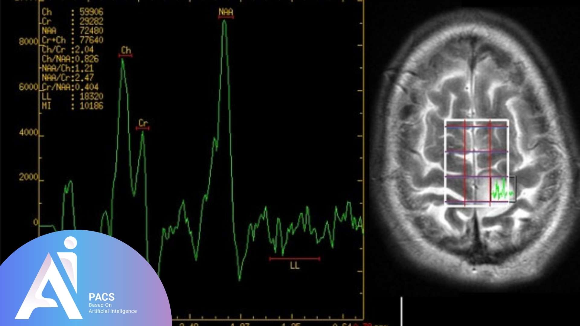
Magnetic Resonance Spectroscopy generates spectra showing peaks corresponding to various metabolites:
- Choline (Cho): Elevated levels may suggest increased cell membrane turnover, often seen in tumors.
- N-acetyl aspartate (NAA): A marker of neuronal health; low levels indicate neuronal loss or damage.
- Creatine (Cr): Reflects energy metabolism and is often used as a reference.
- Lactate: Presence indicates anaerobic metabolism, often associated with tissue injury or tumor hypoxia.
Your radiologist will analyze these peaks and their ratios to differentiate between healthy tissue and disease, aiding diagnosis and treatment planning.
Looking for expert analysis and precise MRI reports? At AI-PACS, we provide accurate and timely online MRI reporting services, offering second opinions from highly skilled radiologists. Whether you’re exploring MR Spectroscopy or need a thorough evaluation of your medical imaging, our team is here to ensure you receive the best care possible. Visit us today for a fast, reliable, and professional MRI report tailored to your needs.
Final Thoughts
Magnetic Resonance Spectroscopy is a powerful tool for metabolic analysis and molecular structure assessment. It provides valuable insights into molecular composition, dynamics, and chemical interactions. This technique is widely used in fields such as chemistry, biochemistry, pharmaceuticals, and medical research, particularly for early cancer detection and disease diagnosis. By analyzing metabolic markers, MRS enhances diagnostic accuracy in neurological disorders, tumors, and metabolic conditions, making it a crucial tool for precision medicine.
References: