

MRI vs. CT Scan: Understanding the Differences and When to Use Each for Accurate Diagnosis
Real-Life Scenario: A Patient’s Journey
Let’s consider Arash, a 45-year-old man who came to the ER after a minor car accident. He had neck pain and a brief loss of consciousness. The ER team immediately ordered a CT scan of the head and cervical spine to check for any fractures or bleeding. Thankfully, the CT showed no acute injury.
Later, because Arash continued to experience numbness in his fingers, his neurologist ordered an MRI of the cervical spine to evaluate the nerves and spinal cord more closely. The MRI revealed a subtle disc herniation pressing on a nerve, something that wasn’t visible on the CT.
This case shows how both imaging tools can work together to provide a complete picture.
What Do These Medical Terms Mean?
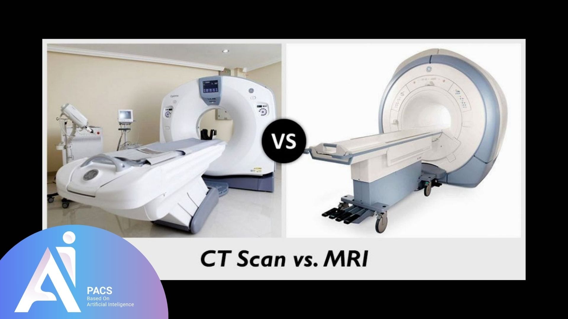
How a CT Scan Works
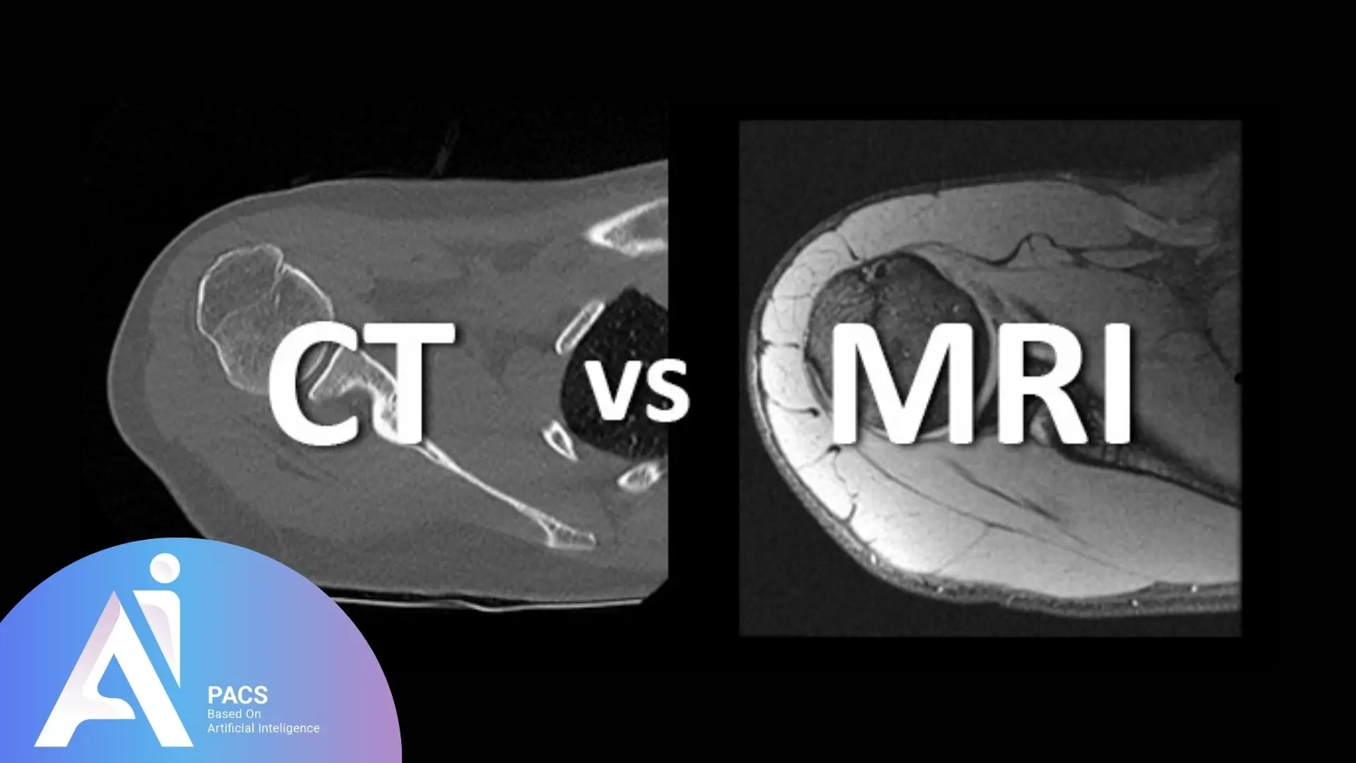
A CT (Computed Tomography) scan uses ionizing X-rays to take quick, detailed cross-sectional images.
The scanner looks like a large ring or donut. You lie on a table that passes through the ring.
Why It Matters: CT is excellent for bones, trauma, chest and abdominal emergencies, and brain hemorrhage.
Duration: Very fast—usually takes just a few minutes.
Radiation Risk: Uses ionizing radiation, which is safe in small doses but not ideal for frequent use.
How an MRI Works
MRI (Magnetic Resonance Imaging) uses strong magnetic fields and radio waves to create detailed images—without using radiation.
MRI machines are tunnel-like. You lie inside a narrow tube, and a device called a coil is placed over the body part being scanned.
You’ll hear loud thumping or knocking noises, which are normal. Earplugs or headphones are usually provided.
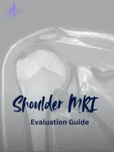
Why It Matters: MRI is best for brain tissue, spinal cord, joints, muscles, and pelvic organs.
Duration: Typically 30–60 minutes per scan.
Safety: No radiation, making it safer for children and long-term monitoring.
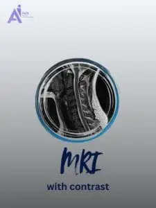
Use of Contrast Agents
Both MRI and CT can use a contrast agent, a special dye injected into a vein to improve image clarity.
CT Contrast: Highlights blood vessels, tumors, and infections.
MRI Contrast: Especially helpful for viewing tumors, inflammation, or nerve-related diseases.
Safety Note: Reactions are rare but possible. Always inform your doctor about allergies or kidney problems.
Which Scan Is Better for What?
MRI Is Preferred For:
- Brain and spinal cord (e.g., multiple sclerosis, tumors, disc herniations)
- Muscles, tendons, ligaments (e.g., sports injuries)
- Pelvic organs (e.g., uterus, prostate)
CT Scan Is Preferred For:
- Head trauma and bleeding
- Bone fractures
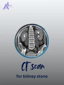
- Chest and abdomen (e.g., pneumonia, appendicitis, kidney stones)
- Emergency situations due to its speed
Noise, Comfort, and Claustrophobia
CT is quick, quiet, and open; better for people with claustrophobia.
MRI is noisier and more enclosed, but staff can provide comfort measures like music or sedation if needed.
The choice between CT and MRI often depends on your specific symptoms and the urgency of your condition. Understanding which scan was chosen for your situation and what it reveals requires expertise in both imaging modalities. Professional interpretation helps you understand why your doctor chose a particular scan and what your results mean for your health.
What Does This Report Say About My Condition?
The type of scan chosen often depends on what your doctor is looking for. For instance:
- If you’re being checked for stroke or trauma, a CT is usually the first step.
- If you’re being evaluated for nerve damage, soft tissue injuries, or brain changes over time, MRI is more sensitive.
Even though MRI and CT use different technologies, their reports can include similar terms—like “mass,” “lesion,” “effusion,” or “herniation.” What matters most is the location, size, and effect on nearby structures—all of which help determine your next step.
If your report seems hard to understand, you’re not alone. An expert review can connect the dots. Whether you’ve had a CT or MRI, expert imaging interpretation provides the clinical context needed to understand your specific findings and their significance for your health and treatment plan.
Why You Should Get an Expert to Review Your Report
Reading an imaging report can feel like reading a puzzle without a picture. Without medical training, it’s easy to misinterpret findings or worry unnecessarily.
That’s why a second opinion from a trained radiologist is so valuable. It gives you:
- A clear understanding of your condition
- Guidance on what findings are important
- Confidence in your next medical step
At AI-PACS, we review both MRI and CT reports and provide detailed explanations that make sense. Whether you’re managing a new diagnosis or just want peace of mind, we’re here to help.
Next Steps: Let’s Help You Understand Your Report
Not sure whether your MRI or CT scan showed something serious? Confused by the medical terms in your report?
If you’re seeking reliable online radiology report services, upload your report now! AI-PACS is here to provide you with the best second opinion on your medical images, delivered by our team of expert radiologists.
Reference:
www.mskcc.org
www.mdanderson.org