


What You Need to Know about a Multiple Sclerosis Brain MRI Report
MS brain MRI report results can seem confusing or even alarming, especially if you’ve had an MRI to evaluate multiple sclerosis (MS) or already have a diagnosis. However, understanding terms like juxtacortical, periventricular, or enhancement after contrast injection is crucial. It puts you in the driver’s seat, helping you interpret what they mean and what they say about the activity or severity of the disease.
This guide breaks down the most common MRI terms associated with MS, allowing you to better understand your report and feel more confident in your care.

What Do These Medical Terms Mean?
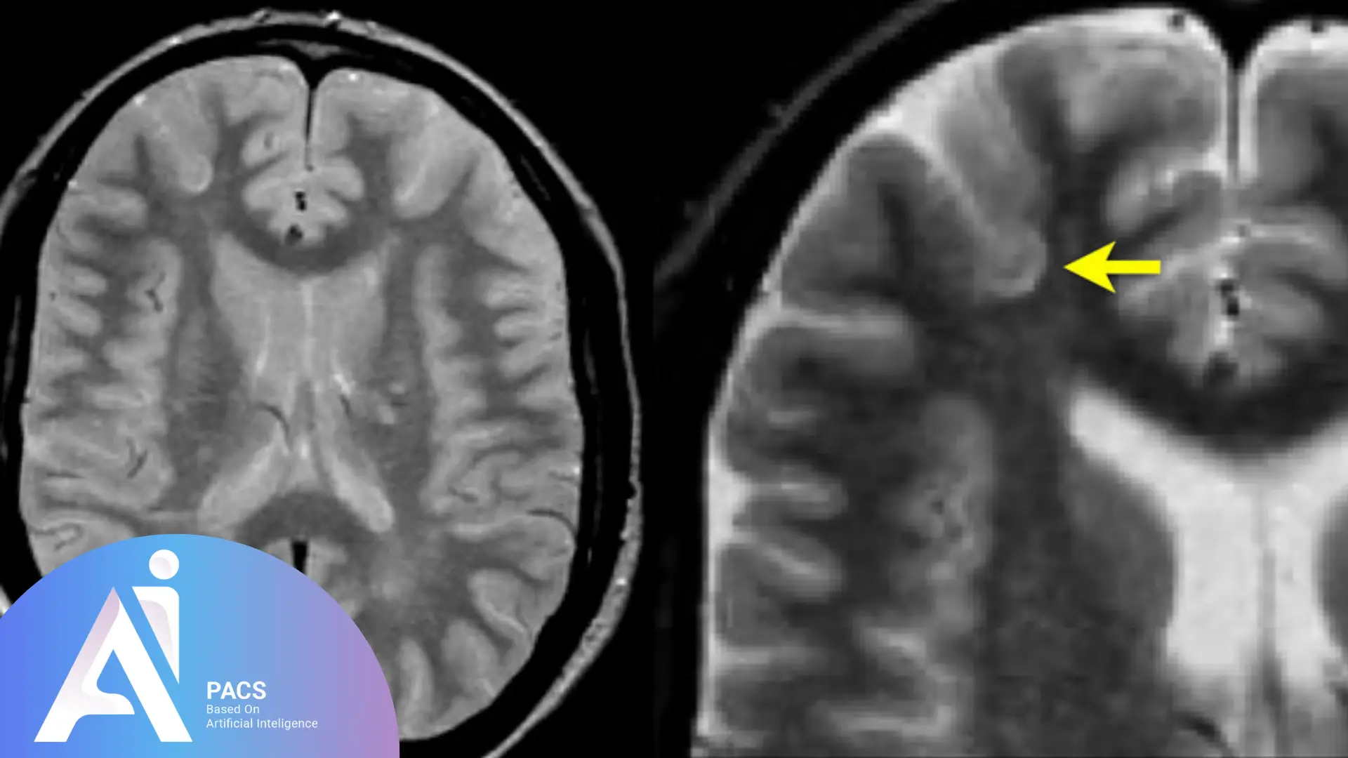
Juxtacortical Lesions
Juxtacortical lesions are areas of damage located near the outer surface of the brain, close to the cerebral cortex. They are often seen in MS patients and are one of the key features doctors look for when diagnosing the disease. These lesions are significant because they reflect the demyelination process, where the protective covering of nerve fibers (myelin) is damaged, disrupting nerve signal transmission.
These lesions are typically one of the first places MS-related damage occurs and are considered a hallmark of the disease. Their presence on an MRI supports the diagnosis of MS and helps clinicians understand the extent of the disease.
Think of the outer layers of your brain as the “processing center” for thought, movement, and sensation. When these areas are damaged by MS, it can lead to problems with motor control, vision, and sensation, making the condition more noticeable.
Periventricular Lesions
Periventricular lesions are found around the brain’s ventricles, which are fluid-filled spaces that help cushion and nourish the brain. These lesions are another key location where MS plaques tend to form.
The presence of periventricular lesions is an important diagnostic feature for MS, as they are commonly seen in people with the condition. They help doctors meet the criteria for MS diagnosis and give insight into the distribution of disease activity in the brain.
These lesions often appear as a ring-like pattern around the ventricles, and their presence is one of the major clues that MS is the underlying cause of the patient’s neurological symptoms.
Enhancing Lesions
Enhancing lesions are areas of damage that appear brighter on an MRI after the injection of a contrast agent (usually gadolinium). This contrast agent highlights areas where inflammation is active in the brain.
The brighter appearance of these lesions indicates that there is active inflammation occurring at the time of the MRI. This suggests that the MS disease is currently active and that the body is experiencing a relapse or a flare-up. Enhancement usually lasts for a few weeks, after which the inflammation subsides.
If you have a lesion that lights up on an MRI scan, it’s similar to shining a flashlight on a dark spot – it shows you where the current damage is happening in real-time, making it easier for doctors to assess disease activity.
New Lesions Compared to Prior MRI
If a new lesion is detected compared to previous MRIs, it may indicate that your MS is progressing or that you are experiencing a relapse. Even if the new lesion does not enhance (appear brighter), its presence can still suggest recent disease activity.
Identifying new lesions helps doctors determine whether the disease is becoming more active or whether your current treatment plan is effective. New lesions suggest a change in the condition that needs to be addressed to manage symptoms and slow progression.
Imagine your MS is like a garden, and each new lesion is a new weed growing. By tracking these lesions over time, doctors can figure out how quickly the disease is advancing and adjust your treatment to manage it better.
Just as MRI can provide critical insights into conditions like multiple sclerosis, it also plays a vital role in Imaging Traumatic Brain Injuries, helping doctors detect and evaluate different patterns of brain damage.
Dawson Fingers
Dawson’s fingers are finger-like projections of damage that extend outward from the brain’s ventricles, often forming a characteristic shape on MRIs of MS patients. These lesions typically follow the path of small veins in the brain.
Named after Dr. James Dawson, who first identified them, these lesions are a highly distinctive feature of MS. Their presence helps confirm the diagnosis, as they are rarely seen in other neurological conditions.
Dawson’s fingers are like “branches” extending from the ventricles, which can be visualized on an MRI and provide a visual clue to doctors about the presence of MS.
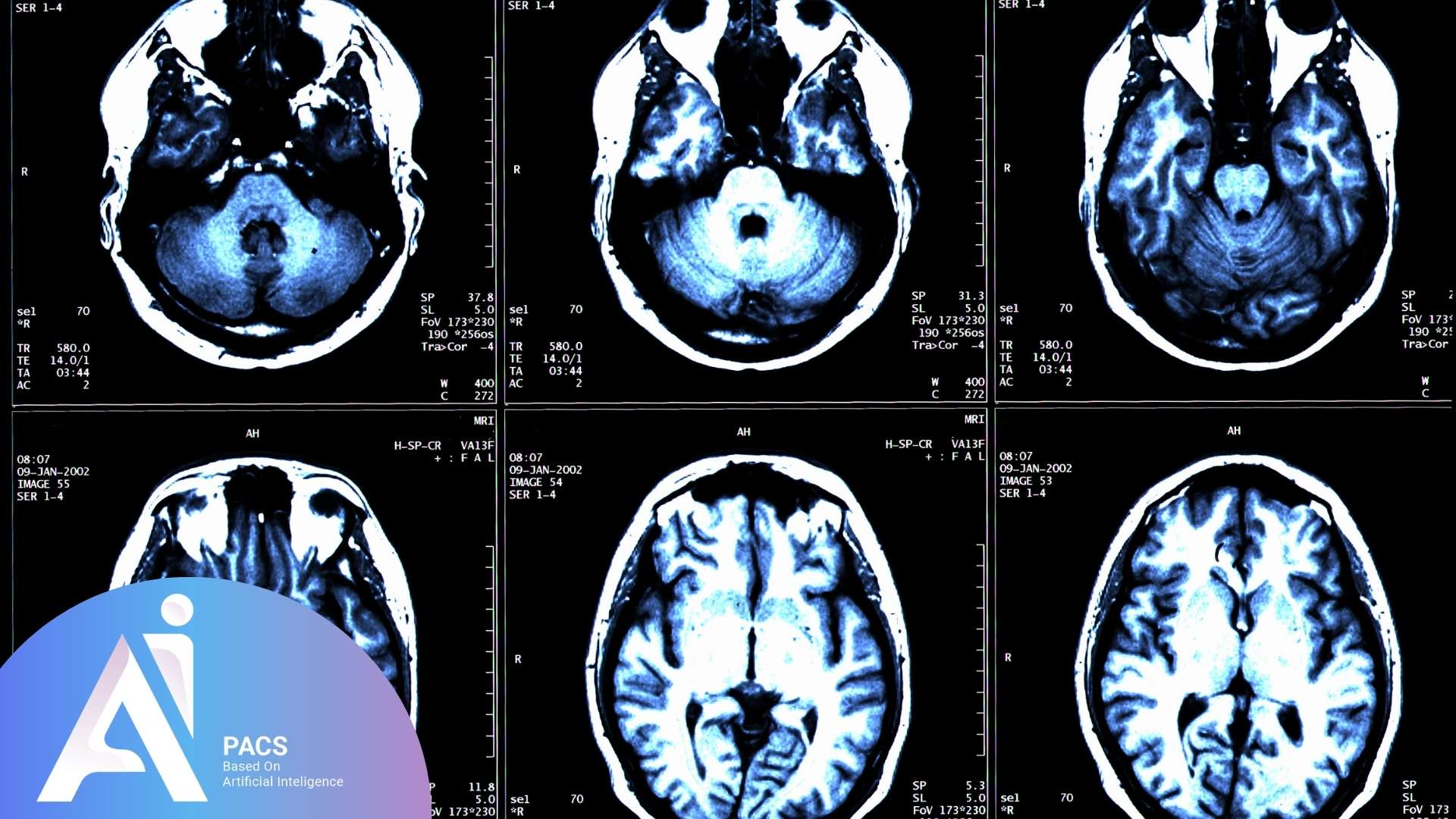
Infratentorial Lesions
These lesions are found in the brainstem or cerebellum, areas located below the tentorium cerebelli (a structure that separates the cerebellum from the rest of the brain). Lesions in these regions can affect critical functions like balance, movement, and coordination.
Infratentorial lesions are particularly important in diagnosing MS because damage to these areas can lead to symptoms like dizziness, vertigo, and difficulty with motor control or facial weakness.
Think of the brainstem as the “control center” for vital functions like breathing, heart rate, and motor coordination. Damage here can affect basic movement and physical balance, which can be severe for MS patients.
T1 Hypointense Lesions (T1 Black Holes)
These lesions appear darker than normal tissue on T1-weighted MRI scans and are often referred to as “black holes.” They are usually a sign of permanent damage to brain tissue, representing areas where nerve fibers have been lost due to long-term disease activity.
T1 black holes indicate chronic damage in the brain that has led to the loss of neurons. These areas are darker because they lack the normal tissue volume that would appear lighter on MRI scans. The more pronounced and persistent these lesions are, the more severe the long-term damage to the brain tissue.
Imagine a “black hole” as a permanent scar left by MS after a relapse. Even if the inflammation has settled, the damage to the brain tissue remains, leading to ongoing symptoms.
Neuronal Loss
Neuronal loss refers to the death or degeneration of nerve cells in the brain, often caused by the inflammatory process of MS.
In MS, inflammation not only damages myelin but can also lead to the loss of neurons, which are responsible for transmitting signals between different parts of the brain and body. Neuronal loss can contribute to permanent symptoms such as weakness, numbness, or cognitive difficulties.
Think of neurons as “wires” that transmit messages in your brain. When these wires are damaged or lost, it leads to slower, disrupted communication, which results in the neurological symptoms of MS.
Brain Parenchymal Atrophy
This refers to the shrinkage or loss of brain tissue, particularly in the areas responsible for cognitive functions and motor control.
Chronic inflammation from MS causes long-term damage to the brain’s neurons and the supporting tissue around them. Over time, this damage leads to a decrease in brain volume, which can be observed as brain parenchymal atrophy on an MRI.
Atrophy is a sign that MS is causing lasting damage to the brain. This shrinkage can result in cognitive decline, memory problems, and difficulty with coordination. Tracking atrophy over time helps doctors monitor disease progression and adjust treatment.
You can think of brain atrophy like the gradual shrinking of a sponge that has been exposed to water over time. As the tissue deteriorates, it becomes less functional, leading to more noticeable symptoms.
What Is the McDonald’s Criteria?
The McDonald’s Criteria is a globally recognized set of diagnostic guidelines used to confirm multiple sclerosis (MS), a disease that affects the central nervous system. MS can be difficult to diagnose because its symptoms often overlap with those of other conditions, and its course can be unpredictable. The McDonald’s Criteria helps doctors make a diagnosis by looking at two critical factors: dissemination in space and dissemination in time. By considering these factors, doctors can confirm whether a person has MS and if the disease is progressing.
Dissemination in Space
“Dissemination in space” refers to the presence of lesions in multiple areas of the brain or spinal cord that are typical of MS. The criteria require that lesions be located in at least two of the following key regions:
- Periventricular: Around the ventricles (fluid-filled spaces in the brain).
- Juxtacortical: Close to the cerebral cortex, which is the outer layer of the brain.
- Infratentorial: Located in the brainstem or cerebellum, areas responsible for balance and coordination.
- Spinal Cord: Lesions affecting the spinal cord are also significant.
For MS to be diagnosed, the lesions must be located in these typical areas where MS plaques are most commonly found. If lesions are seen in just one of these areas, the doctor may need further evidence before confirming the diagnosis. If lesions are seen in multiple areas (e.g., in both the periventricular and spinal cord regions), it strongly supports the diagnosis of MS.
A patient with lesions in both the brainstem and the spinal cord is more likely to have MS than someone with lesions in only one of these areas, as MS typically affects multiple parts of the central nervous system at once.
Dissemination in Time
“Dissemination in time” refers to evidence that the disease has affected the central nervous system at different points in time. MS often presents in a relapsing-remitting pattern, where the disease becomes active, causing new damage, and then later stabilizes or worsens. For MS to be diagnosed, there needs to be:
- Evidence of new lesions that were not present in previous MRIs, indicating that the disease has progressed or relapsed.
- Enhancing and non-enhancing lesions: Enhancing lesions (which appear bright on an MRI after contrast injection) suggest recent or active inflammation. Non-enhancing lesions (which appear darker) represent older damage, showing that the disease has been ongoing for some time.
The combination of old and new lesions on an MRI supports the idea that MS has been active over a period of time, even if the symptoms have not been continuously severe.
A person who has old lesions in the brain from past episodes and new lesions indicating an active relapse could have their MS diagnosis confirmed under the McDonald Criteria. The presence of both types of lesions—enhanced and non-enhanced—indicates the disease has affected the brain over time and is still actively progressing.
How the McDonald’s Criteria Helps with Diagnosis
By combining dissemination in space and dissemination in time, the McDonald Criteria allow doctors to diagnose MS even when symptoms are subtle or intermittent. These criteria enable early diagnosis, which is important because the earlier MS is identified, the sooner treatment can begin to slow disease progression, reduce relapses, and manage symptoms.
For instance, if someone has symptoms like vision problems or muscle weakness but no clear indication of MS, doctors can use MRI findings and the McDonald Criteria to confirm whether MS is the cause. These criteria help distinguish MS from other neurological conditions and provide doctors with the necessary information to begin appropriate treatment.
What Does MS Brain MRI Report Say About My Condition?
If your MRI report includes enhancing lesions, new plaques, or classic features such as Dawson’s fingers, it suggests active MS. But it’s important to know:
Not All Lesions Indicate a Current Relapse
Lesions on an MRI do not always mean you are experiencing a relapse. MS can cause lesions that remain stable over time without causing new symptoms. While enhancing lesions (bright spots after contrast injection) indicate active inflammation, other lesions may be older, stable ones that don’t currently represent new disease activity. Just as scars can remain after an injury, older lesions in the brain can persist without causing new symptoms, even if they’re visible on your MRI.
T1 Black Holes Indicate Older Damage
As we mentioned before, T1 black holes appear as dark spots on MRI scans, indicating areas of chronic damage and neuronal loss. Unlike enhancing lesions, black holes do not show active inflammation but rather represent permanent tissue damage from past disease activity. These areas may have been severely impacted by MS, contributing to long-term neurological symptoms. Imagine a field that has been damaged by a storm, and the grass never grows back. The “bare spots” left behind are like T1 black holes—showing permanent damage from previous relapses.
Lesion Count, Size, Location, and Enhancement Provide Key Clues
When interpreting your MRI, the count, size, location, and enhancement of lesions are essential in determining the severity of your MS. More lesions, especially in critical areas like the spinal cord or brainstem, may suggest more significant disease activity. Larger lesions can cause more severe symptoms, while the enhancement of lesions tells us about the activity level of the disease. Think of a map of your brain, with larger or more numerous lesions indicating more widespread damage. If these lesions enhance (become brighter), it’s like signaling that the disease is actively progressing.
Monitoring changes over time is the key. Comparing scans is essential for tracking progression.
Types of Multiple Sclerosis
- Relapsing-Remitting MS (RRMS): The most common type, characterized by flare-ups followed by periods of remission.
- Secondary Progressive MS (SPMS): A later stage of RRMS where the disease steadily worsens without clear relapses.
- Primary Progressive MS (PPMS): Steady progression of symptoms from the onset without remissions or relapses.
- Progressive-Relapsing MS (PRMS): A rare form of MS with steady progression and intermittent relapses.
- Clinically Isolated Syndrome (CIS): A first episode that may later develop into MS.
- Benign MS: A mild form of MS with minimal disability.
The course of MS can vary widely from person to person, and new treatments have allowed for more effective management of symptoms and disease progression. The exact type of MS one has plays a significant role in how the disease is treated and managed over time.
Why You Should Get an Expert to Review Your Report
MS is a complex disease. An expert can:
- Distinguish MS lesions from other conditions
- Determine activity level and chronicity
- Guide decisions about therapy and follow-up
At AI-PACS, we offer expert second opinions to help clarify your report and ensure nothing important is missed.
Next Steps: Let’s Help You Understand Your Report
If you’ve received an MRI report and are unsure what the MS-related terms mean, we’re here to help you understand it fully.
📁 Submit your report for expert review.
references: