

Understanding Your Second-Trimester Ultrasound Reports
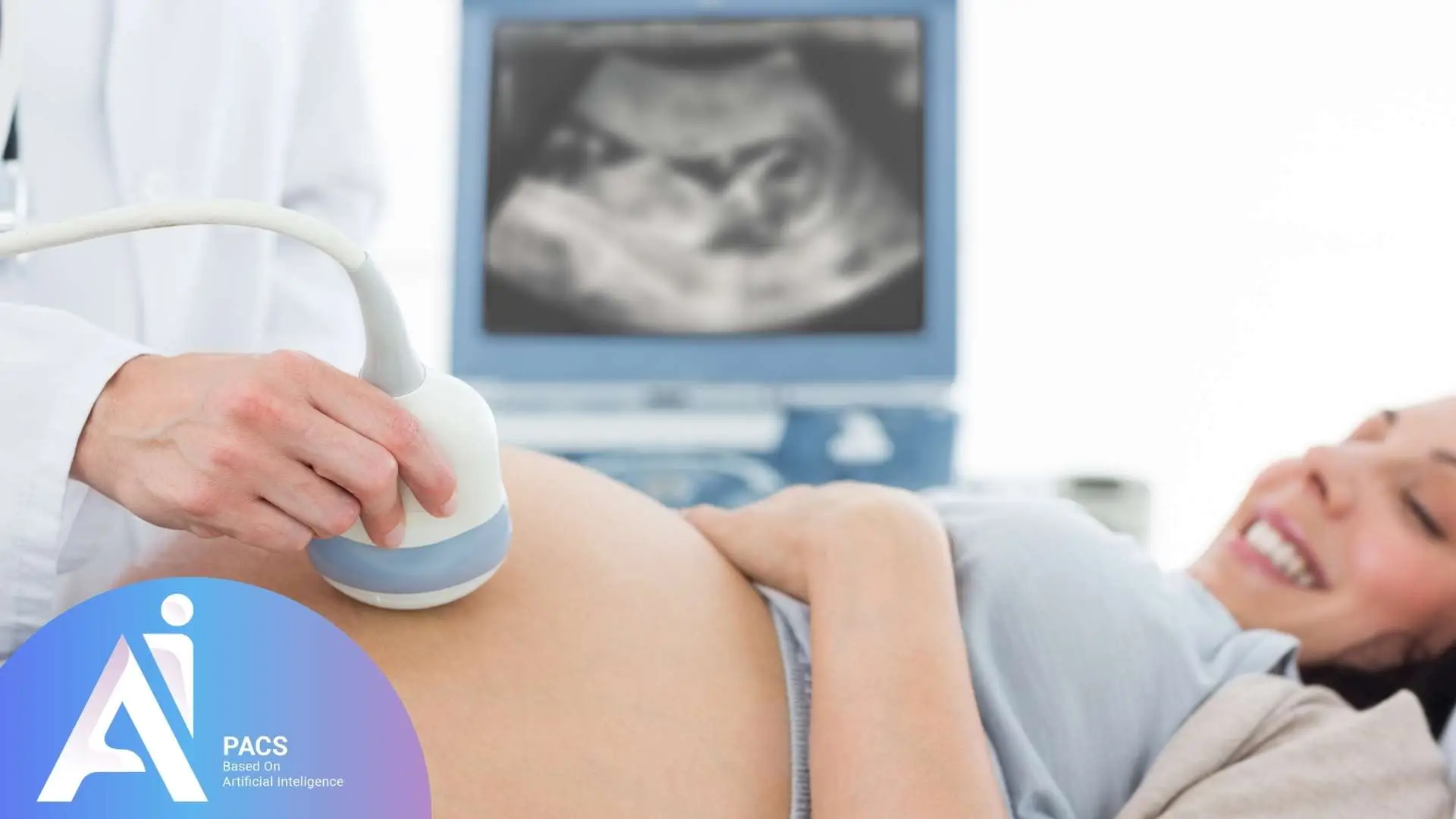
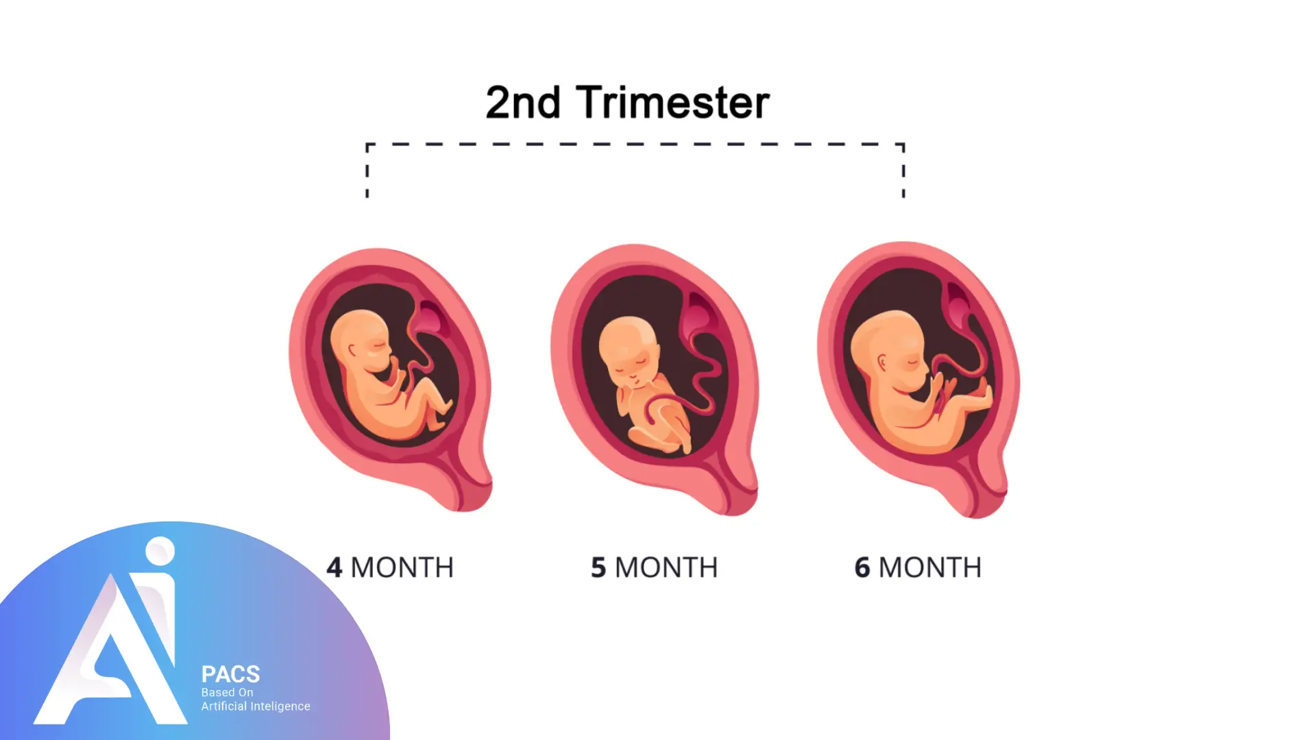
Second-trimester ultrasound in pregnancy reports often contain technical terms and medical abbreviations that can be confusing for many pregnant women. It’s easy to feel overwhelmed. Especially, between months three to six, when a routine scan evaluates the baby’s development, growth, and overall health. This article is designed to help you better understand your second-trimester ultrasound report, providing clear explanations of key findings and guidance on when to seek expert input.
Breaking Down Common Terms in Your Pregnancy Ultrasound Reports
Here are some key terms you might see in your second trimester ultrasound report, along with their plain definitions and why they matter for your pregnancy.
Placenta Location: Understanding Anterior, Posterior, Lateral, and Low-Lying
- Anterior: The placenta is located on the front wall of the uterus.
- Posterior: The placenta is located on the back wall of the uterus.
- Lateral: The placenta is located on the side of the uterus. It can be further specified as:
- Right Lateral: The placenta is on the right side of the uterus.
- Left Lateral: The placenta is on the left side of the uterus.
- Low-Lying: The placenta is near or covers the cervix (also known as placenta previa).
- The location of the placenta plays a critical role in determining how the pregnancy progresses and influences the method of delivery. Most pregnancies involve a posterior or anterior placenta, both of which are considered normal. If the placenta is low-lying or covers the cervix (placenta previa), it may require closer monitoring as the pregnancy advances. In these cases, a cesarean section may be necessary for safe delivery.
- The lateral position of the placenta (right or left) is typically considered normal and does not usually cause issues during pregnancy. However, your doctor will continue to monitor the position of the placenta in subsequent scans to ensure everything is progressing well.
Fetal Presentation: Understanding Baby’s Position
- Fetal presentation refers to the position of the baby in the womb, particularly which part of the body is closest to the cervix.
- Types of Presentation:
- Cephalic: The baby’s head is down.
- Breech: The baby’s feet or bottom are down.
- Transverse: The baby is lying sideways.
- The fetal position in the second trimester is still fluid, and it can change as the pregnancy progresses. By the third trimester, however, the position becomes more important for planning the delivery.
Fetal Movement: Monitoring Baby’s Activity
- This refers to how active the baby is in the womb. Some ultrasounds may capture fetal movements.
- Fetal movement is a good indicator of your baby’s health. A decrease in movement can sometimes indicate a potential issue, so monitoring the baby’s activity can help identify problems early.
What Does AFI (Amniotic Fluid Index) Mean and Why Does It Matter?
- The total amount of amniotic fluid surrounding the baby.
- The depth of fluid pockets in the four quadrants of the uterus is added together.
- Normal Range: 8-24 cm.
Amniotic fluid levels are a key indicator of the baby’s well-being.
- Low AFI (Oligohydramnios): Low fluid levels could indicate potential issues such as fluid leakage or kidney problems in the baby.
- High AFI (Polyhydramnios): High fluid levels may suggest complications like diabetes or fetal swallowing difficulties.
AC (Abdominal Circumference): Measuring Baby’s Belly
- This is the measurement around the baby’s abdomen.
- AC is used to estimate fetal weight and evaluate the growth of the baby. Abnormal measurements could indicate conditions like intrauterine growth restriction (IUGR) or macrosomia (a large baby).
HL (Humerus Length): Measuring Baby’s Arm Bone
- This is the measurement of the length of the baby’s humerus, or upper arm bone.
- Just like the femur, the humerus length can help track the baby’s skeletal development and growth. It’s another marker that provides valuable information for the baby’s overall size.
EFW (Estimated Fetal Weight): How Much Does Baby Weigh?
- The estimated weight of the baby based on various biometric measurements like BPD, HC, FL, and AC.
- EFW is useful in tracking the baby’s growth and can help identify potential concerns, such as whether the baby is growing too slowly (IUGR) or too quickly (macrosomia).
EDC (Estimated Date of Confinement): Your Due Date
- The estimated date when your baby is due to be born, calculated based on measurements of fetal growth.
- Knowing your EDC helps you plan and track the progression of your pregnancy. It also assists healthcare providers in monitoring if the baby is growing on schedule or if any adjustments are needed.
Pctl (Percentile): Tracking Your Baby’s Growth Progress
- This refers to your baby’s growth percentile based on biometric data.
- The percentile shows how your baby’s size compares to others at the same gestational age. For example:
- Between 10th–90th percentile: Normal growth.
- Below 10th: May indicate restricted growth (IUGR).
- Above 90th: May indicate a larger baby (macrosomia).
Cervical Length: Monitoring for Preterm Labor Risks
- The measurement of the length of the cervix.
- A short cervical length can indicate a higher risk of preterm labor. Regular cervical length checks are important for identifying those at risk for premature birth, especially in the second trimester.
What Does This Mean for My Health or Pregnancy?
Second-trimester ultrasounds are designed to provide a detailed look at fetal development. If you have any concerns or questions about your ultrasound report, don’t hesitate to seek expert advice. Most findings like an anterior placenta or slightly different biometry are within normal limits. If something unexpected appears, your provider may recommend additional imaging, such as a level II ultrasound or an MRI, for more details.
Types of Abnormal Results in Prenatal Ultrasound
- Structural Abnormalities
These abnormalities refer to physical changes or malformations in the organs or tissues that can be seen on the ultrasound. For example, the ultrasound may show a problem in the baby’s physical development, such as abnormal limb formation or issues with the brain or heart. In other cases, structural abnormalities can be found in the mother’s body, such as cysts or fibroids that might affect pregnancy. These abnormalities can sometimes be minor, but they may also require closer monitoring or intervention, depending on their severity. - Functional Abnormalities
In some cases, the structure of an organ or tissue may appear normal, but its function is not working properly. For example, in a fetal heart ultrasound, the heart may appear to have no structural issues, but it might not be pumping blood efficiently. This kind of issue can be harder to detect and requires careful monitoring. Functional abnormalities often require additional diagnostic tests to determine the exact cause and to assess the impact on the pregnancy. - Developmental Anomalies
Developmental anomalies are issues that arise during the development of an organ or body part. These conditions are particularly important in prenatal ultrasounds as they relate to how the baby is growing in the womb. Some developmental anomalies can resolve on their own over time, while others might require medical intervention or further monitoring. For example, a baby might show a mild heart defect that could correct itself as the baby grows, or there could be a more severe condition, such as a neural tube defect, that would need immediate care after birth.
Anatomy Checked in the Anomaly Scan
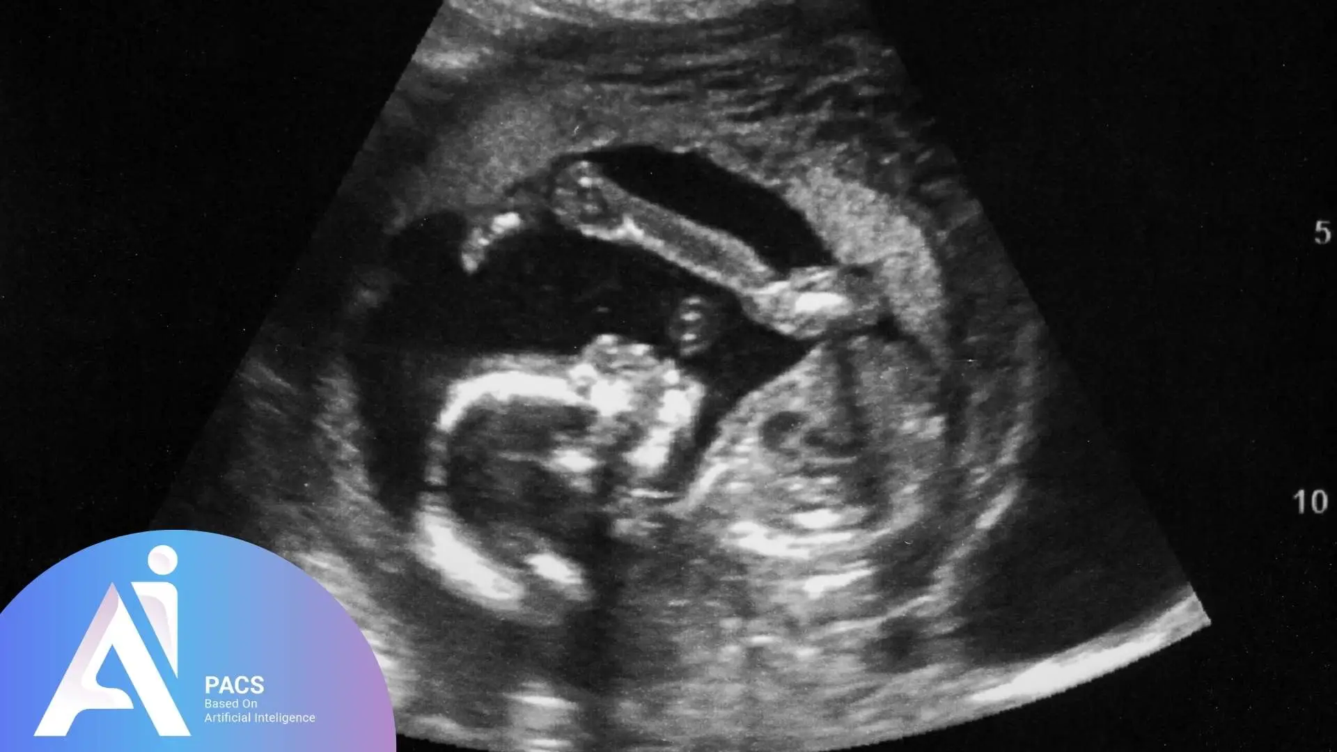
The anomaly scan, typically performed in the 2nd-trimester obstetric ultrasound, provides a comprehensive look at the baby’s development, checking for any structural or functional issues. This process involves carefully examining the fetus from head to toe to ensure everything is developing as it should. Here’s a detailed breakdown of the key anatomical areas and what the sonographer checks for:
- Head and Neck
Starting with the head, the ultrasound checks several key measurements and structures. The biparietal diameter (BPD) and head circumference (HC) are taken to assess the baby’s growth and brain development. The intracranial anatomy, including the cerebellum, posterior fossa, and lateral ventricles, is also surveyed to ensure everything appears normal. These measurements help detect any potential brain abnormalities early on.
For the neck, the ultrasound checks for important markers like the nuchal fold and nuchal translucency. The nuchal translucency is measured earlier in pregnancy (between 11-14 weeks) to detect potential issues like Down syndrome, while the nuchal fold is measured later on (closer to the second trimester). The presence of a nuchal cord, where the umbilical cord wraps around the baby’s neck, is also checked, as it could cause issues during delivery if not monitored properly.
Another area of concern in the neck region is cystic hygromas; fluid-filled sacs that may form around the neck or head. These can indicate problems in the lymphatic system and require further investigation.
- Chest
Moving to the chest, the ultrasound checks the lungs for proper development, ensuring they are the right texture and free from pleural effusion (fluid in the chest cavity) or cystic adenomatoid malformation (a rare lung abnormality). The diaphragm is also assessed to rule out a diaphragmatic hernia, where the diaphragm doesn’t fully close, allowing abdominal organs to enter the chest cavity.
In addition to the lungs and diaphragm, the heart is carefully examined. The four-chamber view of the heart is checked, ensuring all chambers are visible and functioning properly. The aortic arch is also assessed, and the heart’s position in the chest is measured (it should take up about one-third of the chest space, with the apex pointing to the left). The heart rate is measured using M-mode, and any irregularities like arrhythmias, tachycardia (too fast), or bradycardia (too slow) are noted. This helps detect any potential heart conditions early.
- Upper Extremities
The ultrasound will examine the baby’s arms, confirming the presence of both humeri, radius, and ulna. Asymmetries or size differences in the limbs could indicate skeletal dysplasias, which are conditions affecting bone growth. The number of fingers on each hand is also checked to ensure normal development.
- Spine
The spine is imaged in three planes: sagittal, transverse, and coronal. This helps detect any abnormalities such as spina bifida, where part of the spinal cord fails to develop properly. The ultrasound looks for any visible gaps or irregularities along the spinal column.
- Abdomen
Moving to the abdomen, the abdominal circumference (AC) is measured, typically at the level of the stomach and liver. This measurement is essential for estimating the baby’s growth and checking for intrauterine growth restriction (IUGR). The liver is also examined to ensure proper development and to check for hemangiomas (benign tumors), cysts, or calcifications. Gallstones may occasionally be seen, though they are rare.
The ultrasound also checks for abdominal wall defects, such as omphalocele or gastroschisis, where the intestines or other organs protrude through a hole in the abdominal wall.
- Kidneys
The kidneys are examined for their location and echogenicity (how they appear on the ultrasound). Any signs of hydronephrosis (swelling due to urine buildup), renal agenesis (absence of a kidney), or cysts are noted. The kidneys are typically imaged in sagittal, coronal, and transverse planes to get a complete view.
- Cord Insertion
The umbilical cord is checked to ensure it has the usual three vessels: two arteries and one vein. This is important because any variation in the number of vessels could suggest potential issues, such as omphalocele or bladder extrophy.
- Bladder
The bladder is also examined, as it may fill and empty regularly. It’s checked for signs of bladder extrophy, a condition where the bladder develops outside the body, or for a keyhole bladder, which can indicate a blockage in the urinary tract known as posterior urethral valves.
The gender of the baby is often determined at this point as well. The ultrasound can clearly show whether the baby is male or female based on the genital structures.
- Lower Extremities
The femur length is measured, as it’s a key indicator of fetal growth. The length of the femur helps estimate the baby’s overall size, and any discrepancies could point to skeletal dysplasias or growth restriction. The tibia and fibula are also assessed for development.
The baby’s feet are carefully examined to ensure there are five toes on each foot and to check for conditions like club foot, rocker bottom foot, or sandal gap, which could indicate developmental issues.
This comprehensive scan covers the fetus from head to toe, helping to detect any potential problems early in pregnancy. Early detection of abnormalities allows for better management of the pregnancy, providing the opportunity for additional testing, monitoring, or even interventions if necessary. Understanding these checks and their implications is essential for ensuring your baby’s health throughout the pregnancy.
Next Steps After Your Second-Trimester US
After discussing your 2nd-trimester sonography results with your healthcare provider, they will explain the next steps. This might involve additional imaging tests, such as a more detailed ultrasound or an MRI, depending on the findings. Your provider will also consider your overall health, symptoms, and family medical history when deciding on the best course of action.
Understanding your ultrasound results is the first step in making informed decisions about your pregnancy. While it may feel overwhelming at first, your healthcare team is there to guide you through interpreting the results and planning any necessary follow-up care. Asking for a copy of your report can help you prepare questions and better understand what’s next, ensuring you stay actively involved in your care for greater peace of mind. If you want to know about pregnancy doppler ultrasound and understand more you can click on it.
Why Getting a Second Opinion Is a Smart Move
Pregnancy imaging involves profoundly personal and sometimes time-sensitive decisions. Misreading or misunderstanding a report can lead to stress or unnecessary interventions. A second opinion from a women’s imaging expert can help you:
- Gain peace of mind
- Understand what your results really mean
- Decide on appropriate next steps
Need to Help Understand Your Ultrasound Report?
Understanding your pregnancy report is crucial for making informed decisions about your health. As a radiologist with specialized training in women’s and obstetric imaging, I’m here to guide you through your second-trimester ultrasound reports, decoding each medical term for you. If you’ve had a scan related to pregnancy, fertility, or gynecologic health and feel unsure about the report, we’re here to help.
📁 Upload your ultrasound report for expert review
References:
my.clevelandclinic.org
drnadelman
sonographictendencies
bornontario