Nose Job or Nasal Reconstruction CT Scan: What You Need to Know
Article Overview
Why CT Scans Matter Before Nasal Surgery
CT scans play a crucial role in the success of nasal surgeries, whether for cosmetic or functional purposes. They provide detailed imaging of the nasal structure, including bones, cartilage, and soft tissues, helping surgeons plan precise procedures. By identifying underlying issues like a deviated septum, sinus problems, or previous trauma, CT scans minimize the risk of complications and improve surgical outcomes. They are particularly important for patients undergoing reconstructive surgery after injury or those with breathing difficulties, ensuring both aesthetic and functional results.
What is a Nasal Reconstruction CT Scan?
A nasal reconstruction CT scan is a specialized imaging technique that provides detailed, cross-sectional views of the nasal bones, cartilage, and surrounding structures. It uses X-ray technology and advanced computer processing to create 3D images, offering a comprehensive look at the internal anatomy. This scan is essential for planning cosmetic and functional nasal surgeries, ensuring precision and minimizing risks.
Purpose of a CT scan for nasal procedures
The primary purpose of a CT scan for nasal procedures is to give surgeons a detailed roadmap of the nasal anatomy. It helps identify structural issues, such as:
- Deviated septum
- Fractures or nasal bone abnormalities
- Obstructions in the airway or sinuses
- Complications from previous surgeries
These insights allow surgeons to tailor the procedure to the patient’s needs, whether improving aesthetics or resolving breathing problems.
How it differs from other imaging techniques
CT scans differ from other imaging methods like X-rays or MRIs in several ways:
- Resolution: CT scans provide high-resolution images with more detail than standard X-rays.
- 3D Visualization: Unlike X-rays, which show only two-dimensional views, CT scans generate 3D images, which is crucial for complex surgeries.
- Soft Tissue Imaging: While MRIs are better for soft tissue, CT scans balance imaging of both bone and cartilage, making them ideal for nasal procedures.
Typical uses in rhinoplasty and nasal reconstruction
CT scans are commonly used in:
- Cosmetic Rhinoplasty: To assess the underlying structure and guide reshaping.
- Functional Nasal Surgery: To address breathing issues caused by structural defects or sinus blockages.
- Trauma Reconstruction: To evaluate damage after injuries and plan repairs.
- Revision Surgeries: Identifying and correcting complications from previous nasal procedures.
When Do You Need a CT Scan Before a Nose Job?
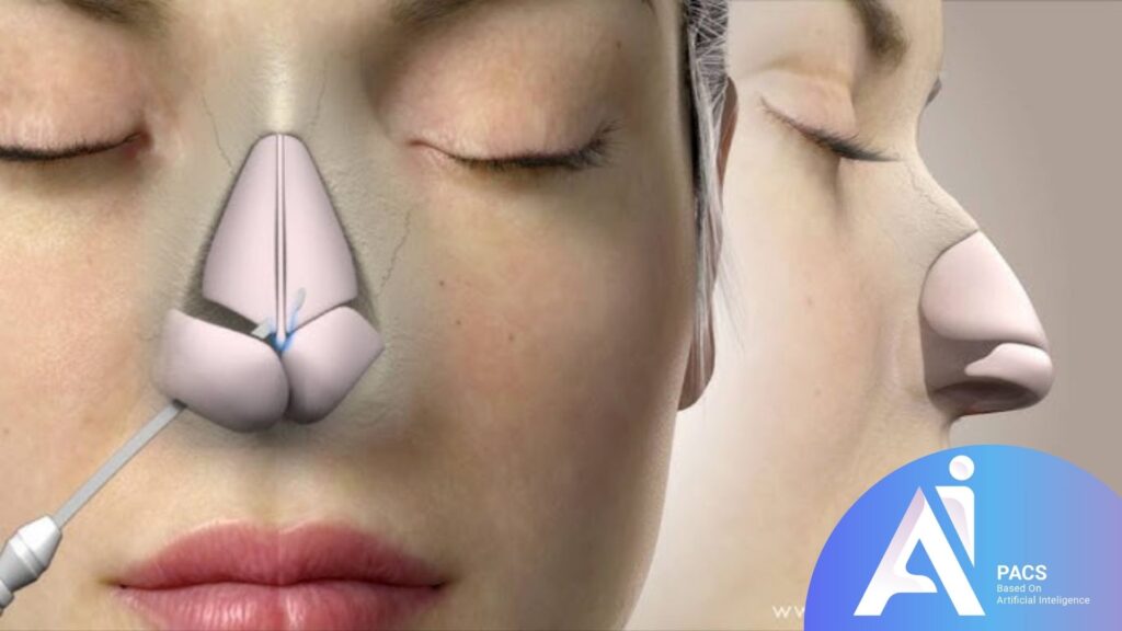
A CT scan is often recommended as part of the preparation for a nose job, depending on the type of procedure and the individual patient’s needs. It provides valuable insights into the nose’s internal structure and surrounding areas, helping to plan a safe and effective surgery. Here are the most common situations where a CT scan is needed before a nose job:
- For Functional Problems: If breathing difficulties, snoring, or obstructed airflow are among the concerns being addressed, a CT scan can help identify issues like a deviated septum or nasal valve collapse.
- After Nasal Trauma: A CT scan can thoroughly evaluate any recent or past injury to the nose that may have caused fractures or structural deformities.
- When Sinus Issues Are Present: Chronic sinus infections, polyps, or blockages often require a CT scan to ensure a comprehensive approach to treatment.
- For Revision Surgeries: If you’ve had previous rhinoplasty and need adjustments, a CT scan reveals any scar tissue or internal changes from earlier procedures.
- Surgeon’s Recommendation: In some cases, surgeons may suggest a CT scan to better visualize anatomical nuances, even for cosmetic procedures.
How is a CT Scan Performed for Nasal Reconstruction?
A CT scan for nasal reconstruction is a quick and non-invasive procedure that provides detailed images of the nasal structures. Here’s how the process typically works:
- Preparation: Patients may be asked to remove any metal objects, such as jewelry or glasses, as these can interfere with imaging. No other special preparation is usually required unless specified by the radiologist.
- Positioning: The patient lies on a motorized table that slides into the CT scanner. For nasal imaging, the head is positioned carefully to ensure accurate results.
- Scanning Process: The scanner rotates around the head, using X-rays to capture cross-sectional images of the nasal area. The process is painless and takes only a few minutes.
- Advanced Technology: Modern CT scanners use low radiation doses while maintaining high image quality, making the procedure safe and efficient.
- Completion and Results: After the scan, the radiologist reviews the images and prepares a report for the surgeon. The entire process typically takes less than 30 minutes.
What Does a CT Scan Reveal?
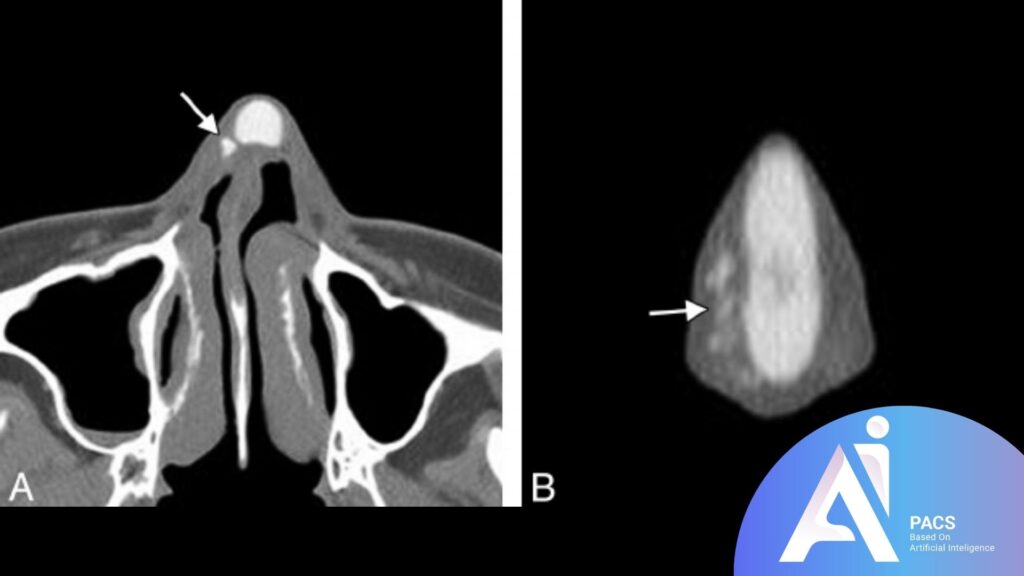
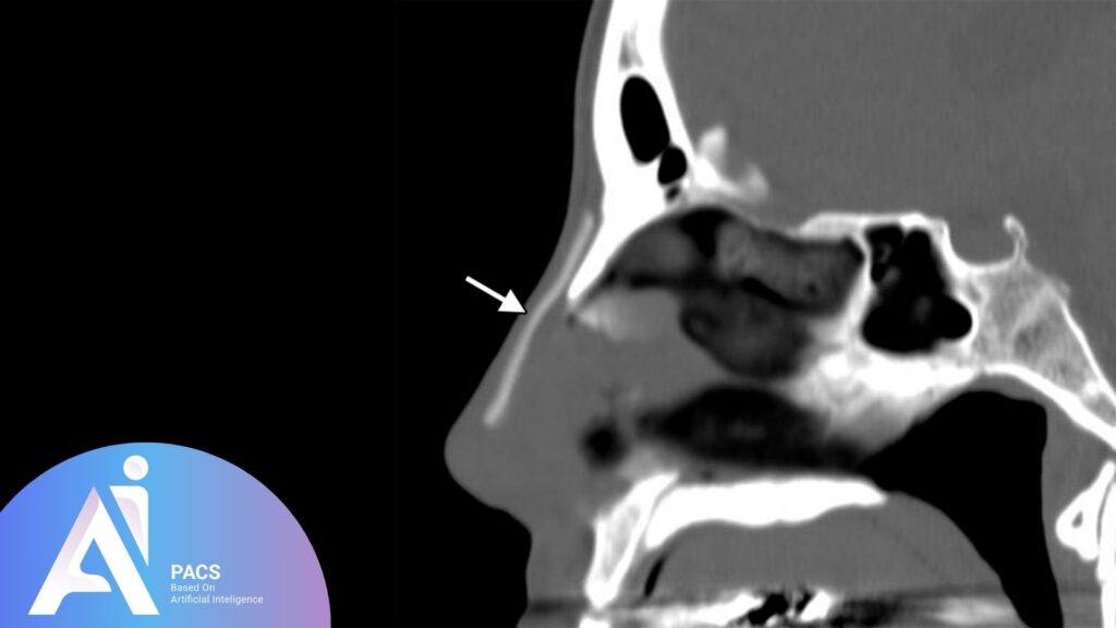
A CT scan offers an unparalleled view of the internal nasal anatomy, providing crucial information for surgical planning. It identifies typical structures and potential abnormalities that could impact the procedure’s outcome.
Key anatomical details captured
Septum: Evaluate the alignment and condition of the nasal septum, highlighting deviations or perforations.
Nasal Bones: Reveals fractures, deformities, or abnormalities in the bony structure of the nose.
Sinuses: Provides a clear view of the sinuses, detecting blockages, polyps, or other issues.
Cartilage and Soft Tissues: This shows the condition of cartilage and surrounding soft tissues, which are essential for cosmetic and functional surgeries.
Airway Pathways: Identifies obstructions or narrow passages affecting airflow.
Identification of abnormalities
A CT scan can detect a wide range of issues that may not be visible during a physical examination, including:
- Deviated septum
- Nasal fractures or bone displacements
- Chronic sinus infections or inflammations
- Nasal polyps or tumors
- Scar tissue or complications from previous surgeries
- Congenital defects affecting nasal structure or function
Benefits of a CT Scan Before Rhinoplasty
Undergoing a CT scan before rhinoplasty offers significant advantages, both for the surgeon and the patient. It provides critical insights into the nasal anatomy, improving the precision of the surgery and minimizing potential risks. Below are the key benefits:
Enhanced precision in surgery
A CT scan allows surgeons to:
Visualize the Nasal Anatomy: Detailed 3D images of the nasal bones, septum, cartilage, and surrounding tissues help create a precise surgical plan.
Address Functional Issues: Identifying structural problems, such as a deviated septum or sinus blockages, ensures that cosmetic and functional concerns are addressed simultaneously.
Custom Tailored Approach: The surgeon can plan exact modifications with detailed imaging, ensuring the changes align with the patient’s desired aesthetic and functional outcomes.
Identify Hidden Problems: CT scans can uncover underlying conditions that might not be visible through external examination, such as undetected fractures or scar tissue from prior surgeries.
Reduced risk of complications
Using CT imaging before surgery helps to:
Avoid Unforeseen Challenges: By knowing the exact structure of the nasal anatomy, surgeons can prevent complications during the procedure.
Minimize Post-Surgery Issues: Addressing structural abnormalities in advance reduces the likelihood of problems like persistent breathing difficulties or asymmetry after surgery.
Improve Healing and Recovery: Precise planning ensures minimal disruption to surrounding tissues, leading to faster recovery and fewer postoperative complications.
Enhance Patient Safety: The detailed information provided by a CT scan ensures the procedure is carried out with the utmost care, reducing risks associated with blind surgical techniques.
Risks and Considerations of CT Scans
While CT scans are valuable for planning nasal surgeries, they have certain risks and considerations. One primary concern is radiation exposure, which, though minimal in modern CT scanners, may accumulate with repeated scans. This is particularly relevant for younger patients or those with a history of frequent imaging. Additionally, if contrast dye is used to enhance imaging, there is a small risk of allergic reactions, ranging from mild skin irritation to more severe responses. Patients with allergies or kidney issues should inform their doctor beforehand.
Cost and accessibility can also be considerations, as CT scans may not always be covered by insurance and can be expensive. Furthermore, while the procedure is quick and non-invasive, some individuals may experience anxiety or claustrophobia during the scan. Despite these potential drawbacks, CT scans remain a safe and effective option for pre-surgical planning when used appropriately, ensuring better outcomes and minimizing surgical risks.
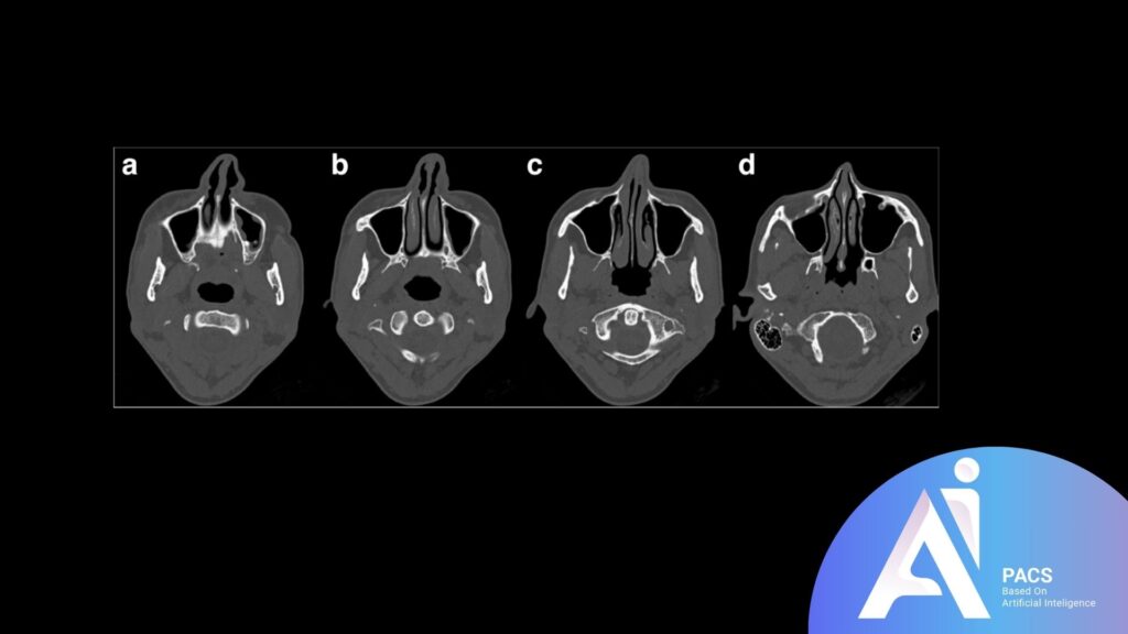
CT Scans for Revision Rhinoplasty
Revision rhinoplasty, or secondary nasal surgery, often involves addressing complex issues left unresolved or caused by a previous procedure. These surgeries require a higher level of precision due to changes in the nasal anatomy, such as scar tissue, altered cartilage, or bone deformities. CT scans play a vital role in these cases by providing a comprehensive view of the internal structures, enabling surgeons to plan the surgery more accurately and ensuring optimal functional and aesthetic outcomes.
Why imaging is critical for secondary procedures
Imaging is essential for revision rhinoplasty because it helps surgeons understand the unique anatomical changes that result from previous surgery. CT scans reveal critical details such as scar tissue, bone misalignment, or cartilage loss that may not be visible during a physical examination. Additionally, they allow surgeons to identify complications like nasal obstructions, collapsed valves, or poorly healed fractures. This detailed understanding ensures precise planning, reduces the likelihood of surprises during surgery, and improves the overall safety and success of the procedure.
Addressing structural issues from previous surgeries
CT scans are invaluable in diagnosing and addressing structural problems caused by prior nasal surgeries. They help surgeons visualize scar tissue and its extent, enabling strategies to minimize its impact. Using the insights from CT imaging, misaligned bones or inadequate cartilage support can also be identified and corrected effectively. Moreover, the scans assist in addressing functional concerns, such as breathing difficulties caused by obstructions or deviations, ensuring that the revision rhinoplasty restores both form and function.
Conclusion
A CT scan is an invaluable tool in planning both primary and revision rhinoplasty, providing surgeons with a detailed and accurate understanding of the nasal anatomy. From identifying structural abnormalities to addressing complications from previous surgeries, CT imaging ensures precision, reduces surgical risks, and enhances patient outcomes. While there are some risks and considerations, such as radiation exposure and cost, the benefits far outweigh these concerns, making CT scans a critical step in achieving safe and successful nasal surgery results. Always consult with your surgeon to determine if a CT scan is necessary for your procedure.


Leave a Reply