What is the TFCC in Wrist MRI Scans: A Comprehensive Guide
The wrist, a marvel of anatomical engineering, strikes a delicate balance between flexibility and stability. The resulting pain and limitations can be significant when issues arise within this complex joint. One often overlooked structure that is crucial for wrist function is the triangular fibrocartilage complex (TFCC). Understanding this key wrist component and its role in wrist injuries is crucial, especially with modern diagnostic techniques such as MRI scans. This article explores the anatomy of the TFCC, its function, the role of MRI in diagnosing injuries to this stabilizing complex, and how patients can seek treatment and recovery.
Overview of Wrist Anatomy
The wrist comprises small bones, ligaments, tendons, and cartilage that work together to allow various movements like flexion, extension, pronation, and supination. The carpal bones connect the hand to the forearm bones, the radius, and the ulna. Located at the intersection of these bones is the fibrocartilaginous part of the wrist, which is crucial for stabilizing the distal radioulnar joint (DRUJ) and supporting weight-bearing activities. Any damage to this system can limit the wrist’s mobility and strength.
Importance of MRI in Wrist Injury Diagnosis
Magnetic Resonance Imaging (MRI) is essential for diagnosing wrist injuries. It provides detailed visualization of soft tissues such as ligaments, tendons, and cartilage, which are not visible on standard X-rays. This is especially important for the wrist support complex, a structure composed of fibrocartilage and ligaments, as even small tears can cause long-term wrist pain. MRI scans can precisely identify these tears, aiding in treatment decisions.

Understanding the TFCC
What is the TFCC?
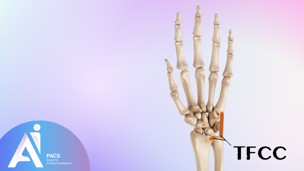
The triangular fibrocartilage complex is a group of
ligaments, tendons, and cartilage located on the ulnar side of the wrist.
It acts as a cushion between the end of the ulna bone and the small bones of the
wrist, providing stability and allowing for smooth movement during rotation and
when bearing heavy loads. Damage to this ulnar-side wrist structure can
lead to ulnar-sided wrist pain, clicking, and instability.
Role of the TFCC in Wrist Stability and Movement
- This key wrist component stabilizes the distal radioulnar joint (DRUJ)
- enabling the wrist to rotate and perform intricate movements.
- It also serves as a shock absorber during weight-bearing tasks like lifting, pushing, or pulling.
If the wrist stabilizing structure is compromised, it can upset this balance, resulting in pain, weakness, and reduced wrist function.
What is the TFCC in Wrist MRI?
How MRI Helps Visualize the TFCC
MRI technology provides high-resolution images that enable
physicians to examine the wrist’s stabilizing complex in fine detail. Since
this structure comprises soft tissue that cannot be seen on regular X-rays, MRI
is considered the best method for assessing its condition. The scan can reveal
tears, degeneration, or inflammation that may be causing pain or instability.
Radiologists use multiple imaging sequences to identify abnormalities that
might otherwise go undetected.
Common Conditions Affecting the TFCC
Tears in the triangular fibrocartilage complex are a
frequent problem, particularly among athletes or people who perform repetitive
wrist movements. Degenerative tears typically happen due to age or overuse,
while traumatic tears are more common in cases of sudden injury, like falling
on an outstretched hand. Other related conditions include inflammation and
ulnar impaction syndrome, where the ulna bone abnormally presses against the fibrocartilaginous part of the wrist.
Anatomy of the TFCC: A Deeper Look
Key Structures Within the TFCC
The triangular fibrocartilage complex consists of several components: the articular disc, the meniscus homologue, the ulnar collateral ligament, and the dorsal and volar radioulnar ligaments. Each of these elements work together to provide stability and support. The articular disc, often referred to as the “central” part of the complex, is a cartilaginous structure that absorbs shock and protects the ulna from direct impact on the carpal bones.How the TFCC Connects to Other Wrist Ligaments and Bones
The wrist support complex is not a standalone structure; it closely interacts with other wrist ligaments and bones. It attaches to the distal radius and ulna and links to the ulnar collateral ligament, which is crucial in stabilizing the distal radioulnar joint. This close relationship means that damage to the wrist stabilizing structure often impacts surrounding structures, magnifying the effects of any injury.Injuries and Tears of the TFCC
Common Causes of TFCC Injuries
A variety of factors can cause injuries to the ulnar side wrist structure. Acute trauma, such as falling on an outstretched hand, can result in a tear. Repetitive strain from activities like tennis, gymnastics, or carpentry can lead to wear and tear over time. In some cases, degenerative changes related to aging can weaken the stabilizing complex of the wrist, making it more susceptible to injury.Symptoms Indicating TFCC Damage
Pain on the ulnar side of the wrist, especially during twisting or gripping movements, is the main symptom of an injury to the fibrocartilaginous part of the wrist. Other symptoms include a clicking or popping sensation, weakness, and a feeling of wrist instability. Swelling and tenderness may also occur, especially after physical activity.How MRI Scans Detect TFCC Injuries
The Process of an MRI Scan for the Wrist
The MRI scan process is non-invasive and typically takes 30 to 45 minutes. During the scan, the patient’s wrist is positioned in the scanner, and multiple sequences are taken to capture different tissue contrasts. Specialized techniques, such as proton-density-weighted imaging, are often used to enhance visualization of the wrist stabilizing structure.
Key Indicators of TFCC Damage in MRI Scans
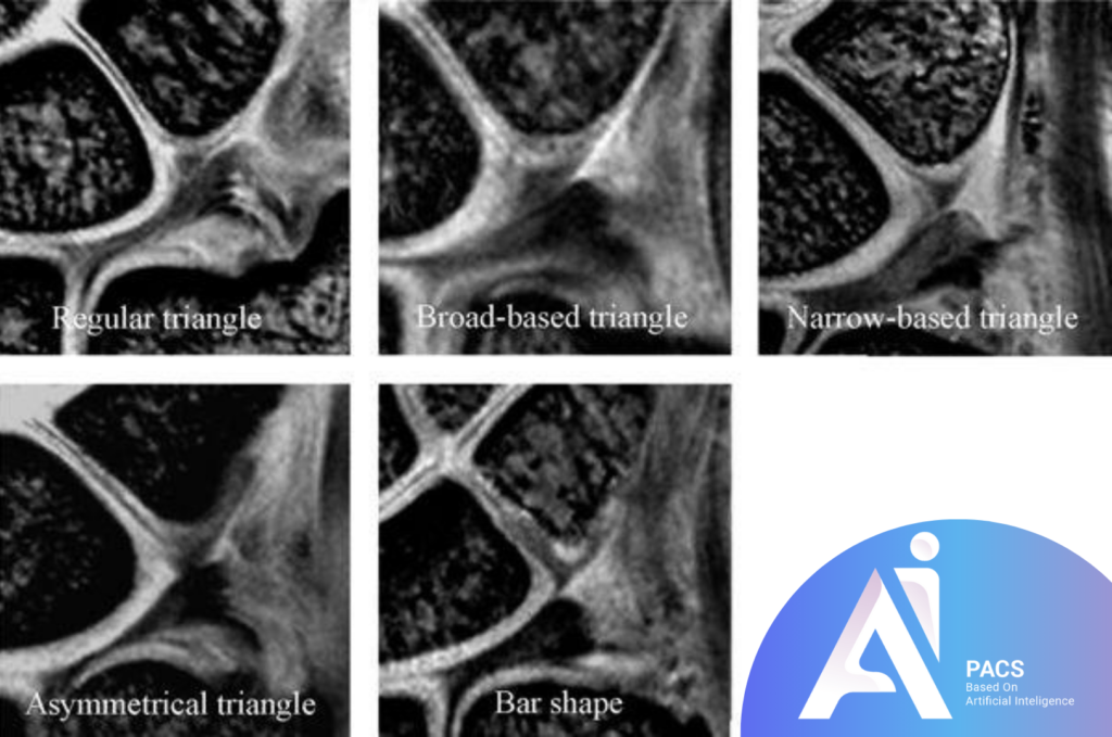
MRI scans can detect various abnormalities within the triangular fibrocartilage complex, such as fraying, partial or full-thickness tears, and degeneration. Radiologists examine changes in signal intensity within the structure to identify tears or abnormalities. The presence of fluid in or around the ulnar-side wrist structure may also indicate damage.

Types of TFCC Tears Seen in MRI
Degenerative vs Traumatic Tears
Degenerative tears of the wrist support complex develop gradually over time and are often associated with aging or chronic overuse. These tears are typically observed in older individuals or those who engage in repetitive wrist activities. In contrast, traumatic tears occur suddenly due to injury, such as a fall or a sudden twisting motion.
Understanding Partial and Full-Thickness Tears
Partial-thickness tears involve only a part of the stabilizing complex of the wrist and may cause intermittent symptoms. Full-thickness tears involve the entire structure and usually result in more severe symptoms, including chronic pain and wrist instability.
MRI Findings: What to Expect
How Radiologists Interpret MRI Result
Radiologists meticulously examine the MRI images to detect any abnormalities in the fibrocartilaginous part of the wrist. They evaluate the size, location, and extent of any tears and assess the condition of surrounding ligaments and bones. The results are then summarized in a detailed report provided to the patient’s physician for diagnosis and treatment planning.
Common Terms Used in MRI Reports Related to the TFCC
Terms such as “partial tear,” “degeneration,” “synovitis,” and “effusion” are frequently mentioned in MRI reports regarding the triangular fibrocartilage complex. Understanding these terms can help patients better comprehend the nature of their injury and its implications for treatment options.
Treatment Options After TFCC Diagnosis
Non-Surgical Treatments for TFCC Injuries
Injuries to the wrist stabilizing structure can often be treated without surgery by using rest, immobilization, physical therapy, and anti-inflammatory medications. Sometimes, a corticosteroid injection may be suggested to reduce pain and inflammation. Additionally, wrist braces can provide support and protection during the healing process.
Surgical Interventions and Recovery Times
In cases of more severe tears or those that do not respond to conservative treatment, surgery may be necessary. Arthroscopic repair or debridement of the stabilizing complex of the wrist is a common procedure for this condition. Recovery times can vary based on the extent of the injury and the specific surgical technique used but typically range from several weeks to a few months.
Preventing TFCC Injuries
Tips for Wrist Care and Strengthening
Strengthening the muscles in the wrist and forearm can help prevent injuries to the **wrist support complex**. Exercises that improve flexibility and range of motion are also beneficial. Additionally, it’s important to take regular breaks from repetitive wrist activities and use proper techniques when lifting or pushing heavy objects.
Activities That Put Stress on the TFCC
Activities involving twisting motions, such as playing racket sports or lifting heavy objects, can exert significant stress on the ulnar-side wrist structure. Maintaining proper wrist positioning and using protective equipment, like wrist guards, can help decrease the likelihood of injury.

When to See a Specialist
How to Know if You Need an MRI for Your Wrist
If you have persistent pain, swelling, or instability in your wrist, especially on the ulnar side, it might be a good idea to see a specialist. If the injury does not improve with initial treatment or if there is suspicion of damage to the wrist stabilizing structure, an MRI scan may be recommended.Seeking Professional Help for Persistent Wrist Pain
Consulting an orthopedic specialist or hand surgeon is essential if wrist pain persists or worsens. Early diagnosis and treatment can prevent further damage and improve long-term outcomes.Conclusion
Key Takeaways About the TFCC and MRI Scans
The triangular fibrocartilage complex plays a crucial role in wrist stability and function, and injuries to this structure can be debilitating. MRI scans provide a precise and non-invasive method of diagnosing these injuries, offering invaluable insights for treatment planning.
Importance of Early Detection and Treatment
Early detection of injuries to the wrist stabilizing structure through MRI can lead to more effective surgical or non-surgical treatment. Prompt care can prevent further damage and aid in recovery.
Reference:


Leave a Reply