
MRI and MR Arthrography for Shoulder Dislocation: Why, When, Who, and A Patient’s Journey in Contact Sport Athletes
Recurrent shoulder dislocation is a common challenge among athletes in contact and combat sports, including judo, wrestling, karate, and taekwondo. These sports involve high-impact and forceful shoulder movements that can lead to joint instability and repeated dislocation.
Imaging (particularly MRI and MR arthrography) is essential for evaluating the underlying damage and guiding treatment plans.
I’m Dr. Vahid Alizadeh. In this article from the “Why, When, Who” musculoskeletal series, I’ll focus on imaging techniques for athletes with shoulder instability, explaining key findings and how advanced imaging helps in real-world clinical situations.
A Patient’s Journey:
From Frustration to Clarity
A 23-year-old male karate athlete presented with recurrent dislocations of his right shoulder during training over the past year. He had multiple MRIs performed at various centers; however, each yielded inconclusive or conflicting reports. Some suggested a mild labral tear, while others labeled the scans as normal.
Frustrated by persistent symptoms, he was referred for specialized evaluation. After reviewing the prior scans and performing an MR arthrography, the assessment clearly identified a Bankart lesion with an off-track Hill-Sachs defect.
He inquired, “What does off-track mean? What is a Hill-Sachs exactly?” We clarified that his humeral head was engaging incorrectly with the socket due to cartilage and bone loss, which raised his risk of future dislocations
With the diagnosis established, a customized treatment plan was created, which included surgical stabilization followed by sport-specific rehabilitation.
Experiencing repeated shoulder dislocations or unclear MRI results? 📁 Upload your MRI or MR arthrography scan for an expert second opinion.
Why Is Imaging Important for Recurrent Shoulder Dislocation?
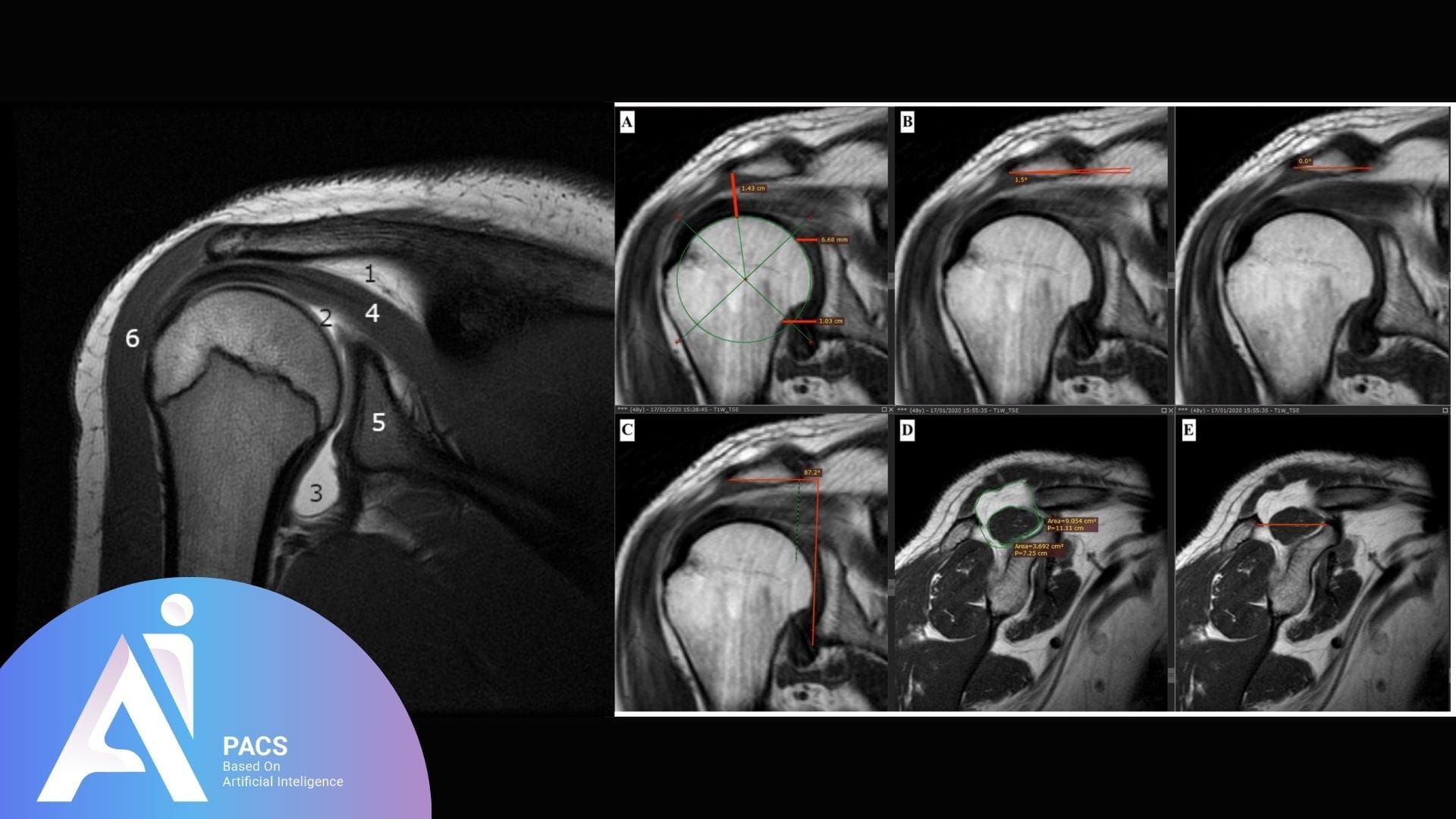
Key Anatomical Structures Affected
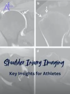
Repetitive dislocations often damage soft tissue and bone structures. Imaging helps visualize:
- Labrum: A ring of cartilage that stabilizes the shoulder socket. Tearing of the labrum, especially in the lower front part (anterior-inferior), is referred to as a Bankart lesion.
- Hill-Sachs lesion: A dent in the back of the humeral head caused when it strikes the socket during dislocation.
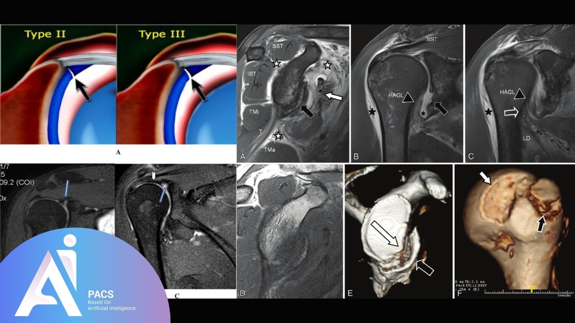
Common Types of Shoulder Lesions
- Bankart lesion: A labral tear without associated bone injury.
- Bony Bankart lesion: A labral tear with an attached bone fragment.
- Hill-Sachs lesion: Compression fracture on the posterior humeral head.
- On-track vs. Off-track lesions: These describe how the Hill-Sachs lesion interacts with the socket. Off-track lesions are more unstable and likely to re-dislocate.
When Is MR Arthrography Preferred Over Routine MRI?
How MR Arthrography Works
- A small amount of contrast dye is injected into the shoulder joint.
- This dye outlines the inside of the joint, revealing tears or loose ligaments more clearly.
- After injection, an MRI is performed as usual.
MR Arthrography vs. Standard MRI
- Labral tears are more visible with contrast.
- Capsular laxity (loose ligaments) is easier to detect.
- Subtle or partial-thickness tears may be missed on MRI but seen on arthrography.
When to Choose MR Arthrography
- Labral or capsular injuries are suspected
- Initial MRI is inconclusive
- Surgical planning is required for high-performance athletes
Who Should Get Shoulder MRI or MR Arthrography?
Ideal Candidates for Imaging
- Athletes in sports with high collision or overhead load (judo, wrestling, boxing, taekwondo)
- Individuals with multiple shoulder dislocations
- Patients with clicking, catching, or slipping sensations in the joint
- Those who have had shoulder surgery and continue to feel unstable
Benefits of Early Imaging
Early imaging leads to:
- Better understanding of injury type
- Tailored rehabilitation programs
- Avoidance of repeated dislocations
- More accurate surgical decision-making
AI-PACS Is With You:
AI-PACS, an advanced imaging system, plays a crucial role in the diagnosis and treatment of shoulder dislocations in athletes. It assists in the interpretation of complex imaging results, ensuring accurate diagnosis and effective treatment planning.
Expert Support for Shoulder Imaging
At AI-PACS.com, we offer comprehensive expert reviews for shoulder MRI and MR arthrography. Our team of experienced radiologists, specializing in sports injuries and joint instability, provide detailed interpretations and recommendations based on the latest medical knowledge and imaging technology.
Our radiologists at AI-PACS.com are well-versed in the patterns of labral damage, bony defects, and capsular issues that athletes face, ensuring you are in good hands.
Need clarity before surgery or rehab?
Need a second opinion on your medical images before surgery or rehab? Get expert analysis from AI-PACS radiologists. Start your online imaging report here.
Final Thoughts
Recurrent shoulder dislocations in athletes often result in damage that can’t be detected by physical exam alone. MRI, especially MR arthrography, provides the clarity needed for accurate diagnosis and treatment.
Whether it’s identifying a Bankart lesion, confirming a Hill-Sachs defect, or planning return-to-sport care, accurate imaging brings a sense of relief and confidence to athletes, helping them recover safely and confidently.
With AI-PACS, you gain access to imaging experts who understand sports medicine and are dedicated to guiding every step of your recovery, providing you with the support and care you need.