


Brain MRI Second Opinion by Expert Radiologists

Accurate, independent review of your brain MRI — typically delivered in under 24 hours

Upload Brain MRI for Second Opinion Team review with a brain neuroradiology expert • Secure upload • Confidential review
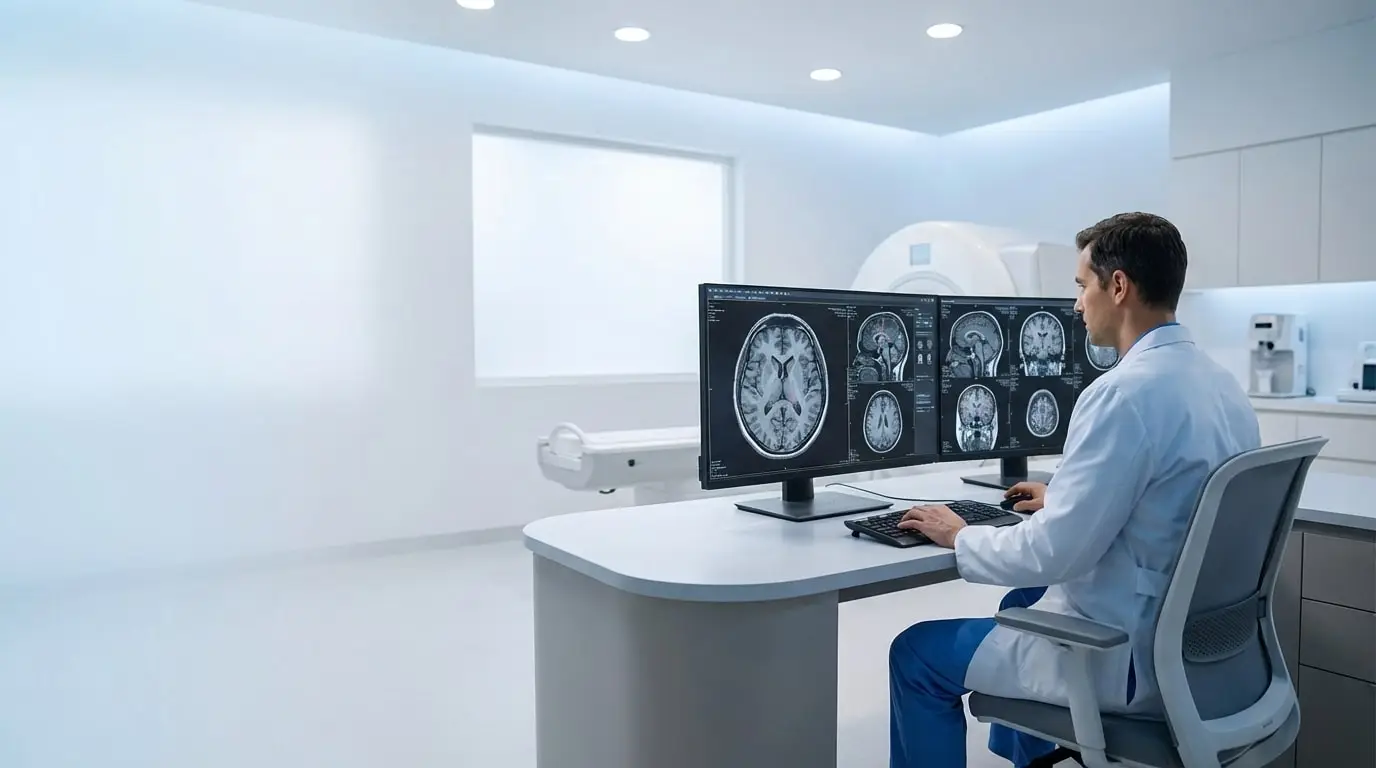
Brain MRI second opinion review by expert neuroradiologists

Why Get a Second Opinion on a Brain MRI?

Interpreting a brain MRI is rarely straightforward. Reports may include terms that sound concerning, yet provide little clarity about their real clinical significance.

“The report mentions a lesion or signal change.”

Small lesions or signal abnormalities are common in brain MRI and may be non‑specific. Without context, it’s often unclear whether these findings are meaningful or incidental.

“White matter abnormalities were noted.”

White matter changes can be related to age, migraine, vascular factors, or other benign causes — but this is not always explained clearly in routine reports.

“I’m unsure if this finding explains my symptoms.”

Patients are often left questioning whether MRI findings truly relate to headaches, dizziness, seizures, or other neurological symptoms.

In many cases, the greatest challenge is not the finding itself, but the uncertainty around its meaning. A brain MRI second opinion can help clarify findings before further clinical decisions are made.

Who Reviews Your Brain MRI Images?

Your brain MRI second opinion is reviewed by a three-person radiology team, including at least one expert neuroradiologist specializing in brain imaging. The team reviews your images, any existing report, and the clinical information you provide. The team lead brings the findings together, prepares the final report, and signs it. Your report is not generated automatically.

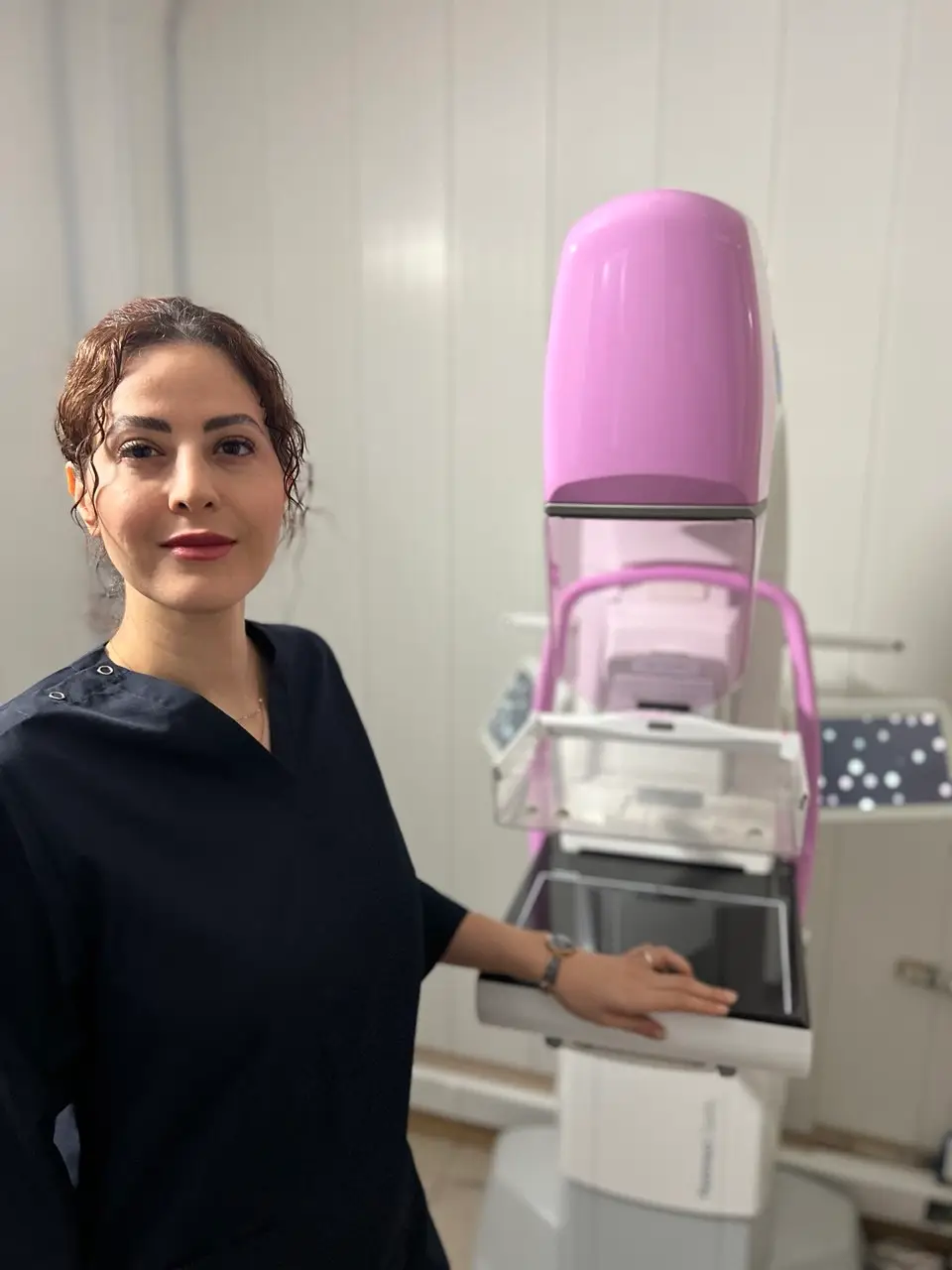
Breast Imaging Radiologist

Dr. Bahare Mobasheri​

Radiologist

Experienced in interpreting brain MRI studies, with a clinical focus on evaluating structural, inflammatory, and incidental intracranial findings.
MC-NO: 133284

Breast Imaging Radiologist

Dr. Vahid Alizadeh

Radiologist

Extensive experience reviewing complex brain MRI cases and providing independent second‑opinion assessments. Member of the European Society of Radiology (ESR) | ESR ID: 160956

Breast Imaging Radiologist

Dr. Reza Alizadeh

Radiologist

Long‑standing experience in interpreting brain MRI examinations, including correlation with clinical history when imaging findings are subtle or non‑specific.
40 Years of Diagnostic Excellence

Our Online Brain MRI Review Process

A simple, secure, and transparent process to receive an expert brain MRI second opinion.

📤

Upload MRI Images

Upload the original DICOM files from your brain MRI. Include the first report if available, your main questions, and any relevant earlier scans. Tell us about changes in symptoms or treatment since the first interpretation. If you need help sharing your files, contact us on WhatsApp.

🔍

Initial Review & Cost Confirmation

A radiologist performs an initial image check to confirm completeness and provides a clear review timeline and cost estimate—typically within 1–2 hours.

💳

Secure Payment

Complete your payment through a secure and encrypted system. Multiple international payment options are available for your convenience.

✅

Expert Report & Follow‑Up Questions

Receive a detailed written brain MRI review. You may also ask follow‑up questions to clarify findings or terminology.

Upload Your Brain MRI

Typical turnaround time: under 24 hours after payment, once complete, readable DICOM images are available.

Conditions We Commonly Review

Brain MRI findings can be diverse and sometimes non‑specific. Our second‑opinion reviews focus on commonly encountered neurological imaging questions, while always considering clinical context and imaging limitations.

White matter abnormalities and signal changes
Suspected demyelinating processes (including multiple sclerosis)
Incidental brain lesions and focal abnormalities
Ischemic changes, small vessel disease, and prior infarcts
Masses, cysts, and space‑occupying lesions
Post‑traumatic findings and microhemorrhages
Congenital variants and developmental abnormalities
Atrophy patterns and age‑related changes
Post‑surgical or post‑treatment imaging changes

These reviews are based on imaging appearance alone and are provided to support — not replace — clinical evaluation by your treating physician.

What your brain MRI second opinion can help clarify

If your report mentions a white matter change, a lesion or another finding that leaves you unsure, tell us what you want clarified. An independent review examines your brain MRI images alongside the relevant medical information you provide.

Your questions might include:

  • “What is visible on my MRI, and what remains uncertain?”
  • “How do these findings relate to the question my clinician is investigating?”
  • “What has changed compared with my earlier brain MRI scans?”

Comparing your current and earlier brain MRI scans

For an Advanced Consultation, share the complete available examinations with their dates and relevant clinical records. Include treatment or surgery dates when they are relevant to your question.

When the studies are suitable for comparison, the review can bring relevant findings together and explain visible changes or similarities across your imaging history. Differences in technique, missing sequences or unavailable earlier images can limit the comparison.

A second opinion may agree with the original interpretation. The review explains what the supplied images can and cannot establish and supports discussion with your treating clinician.

What you receive from a brain MRI second opinion

You receive a written radiology opinion based on your brain MRI images and the relevant medical information you provide. It describes the findings, explains the impression and identifies limitations. After delivery, you can ask questions about the report.

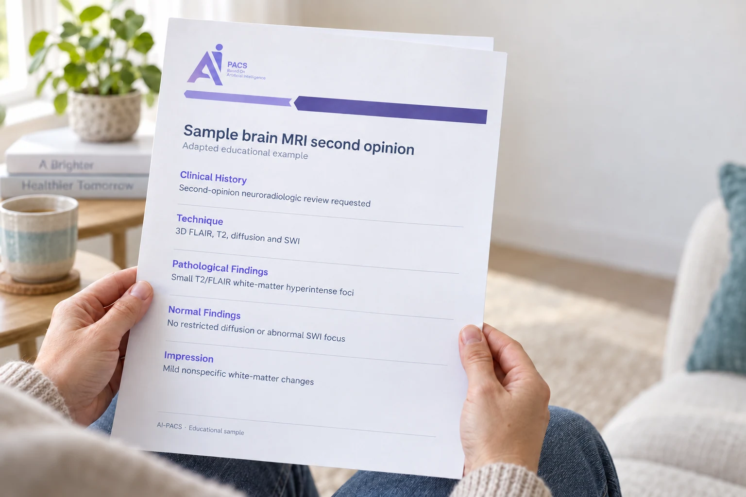
An example of the written opinionThis adapted excerpt illustrates the structure and wording of a brain MRI review. Your own report reflects your images and the questions you ask.
Illustration of an anonymous person holding an AI-PACS sample brain MRI second opinion reportAI-generated illustration · adapted sample
A report you can discuss with your treating teamThe image shows a shortened illustrative layout. Read the adapted report excerpt below.

Inside the sample · 1 of 3

What was reviewed?

Your clinical information and the available MRI sequences

1

Clinical History

Tell us what concerns you and what your clinician is investigating. The written opinion considers the relevant medical information you provide alongside your images.

2

Technique

The review identifies the available MRI sequences and explains important limitations. Missing sequences or differences in technique may affect what can be concluded or compared.

Adapted educational example. Selected report wording has been shortened and identifying details removed. This is not an original patient report or a promised review outcome.
Read the adapted sample brain MRI report

Adapted educational excerpt · identifying details omitted

Clinical History

Second-opinion neuroradiologic review requested.

Technique

Multiplanar, multisequence MRI of the brain was performed before and after intravenous gadolinium administration. Sequences include 3D FLAIR, T2-weighted imaging, diffusion-weighted imaging with ADC maps, susceptibility-weighted imaging (SWI), pre-contrast T1-weighted imaging and post-contrast 3D T1-weighted imaging.

Pathological Findings

Multiple small T2/FLAIR hyperintense foci are present within the white matter of both cerebral hemispheres, predominantly involving the bilateral frontal lobes. Following intravenous contrast administration, none of the described white-matter lesions demonstrates abnormal enhancement.

Normal Findings

No focal intra-axial mass lesion is identified. No focus of restricted diffusion is identified to suggest an acute ischemic lesion. SWI demonstrates no abnormal susceptibility focus.

Impression

The morphology and distribution of these white-matter lesions are not typical for multiple sclerosis or a characteristic demyelinating plaque pattern. Overall appearance is more in keeping with mild nonspecific/chronic small-vessel white-matter changes.

Want an independent review of your brain MRI? Share your complete DICOM images, relevant clinical history and the questions you want clarified. If your question involves changes over time, include the earlier complete examinations with their dates.

Request a brain MRI second opinion

Diagnostic review requires complete DICOM images. The team confirms the scope and cost before payment.

Patient Experiences about Second Opinions Services

Ayesha, Pakistan
Breast MRI Second Opinion
“The second opinion helped me understand whether my diagnosis was accurate.”
Maribel, Mexico
Breast Ultrasound Consultation
“The ultrasound consultation explained why immediate biopsy was not required.”
Tanya, Ukraine
Breast MRI Review
“The MRI findings were explained clearly and without unnecessary alarm.”
Rosa, Brazil
Breast Cancer Second Opinion
“The second opinion supported my doctor’s treatment plan and reduced uncertainty.”
Noura, Egypt
Breast Ultrasound Review
“The ultrasound report was detailed and helped reassure me during follow‑up.”
Priya, India
Mammogram Second Opinion
“The mammogram review helped confirm that further imaging was appropriate.”
Salma, Morocco
Breast Cancer Consultation
“The second opinion helped avoid rushing into unnecessary procedures.”
Marta, Poland
Breast MRI Consultation
“The MRI review explained risk factors in a balanced and understandable way.”
Ana, Peru
Breast Ultrasound Review
“The ultrasound findings were explained clearly and answered my main concerns.”
Ivana, Croatia
Comprehensive Breast Imaging Review
“The overall imaging review ensured no important detail was overlooked.”
Elena, Spain
Breast MRI Review
“The review clarified subtle findings that were confusing in my original report.”
Julia, Argentina
Breast MRI Second Opinion
“The second opinion was detailed and eased my anxiety about the results.”
Lana, Serbia
Breast Ultrasound Consultation
“The consultation gave me confidence that monitoring was the right choice.”
Hana, Jordan
Breast Cancer Imaging Review
“The consultation gave me confidence that monitoring was the right choice.”
Daria, Lithuania
Breast Ultrasound Review
“The report was easy to understand and addressed all my concerns.”
Daniel, Canada
Chest CT Consultation
“The second opinion identified small nodules and explained their low‑risk characteristics in a very clear way. The risk stratification was extremely helpful.”
Sofia, Spain
Musculoskeletal MRI Review (Knee)
“The MRI findings were explained in detail, including ligament integrity and cartilage condition. The report helped my orthopedic surgeon finalize the surgical decision.”
Carlos, Mexico
PET‑CT Oncology ReviewMRI Review (Knee)
“The metabolic activity was analyzed in depth, distinguishing post‑treatment changes from possible recurrence. This distinction was crucial for my oncologist.”

Testimonials are shared with patient permission. Last names and identifying details have been removed to protect privacy.


How Much Does a Brain MRI Second Opinion Cost?

Choose the level of review that best fits your situation. All evaluations focus on accurate brain MRI assessment and clear clinical guidance.

Basic Plan
A professional written second‑opinion report focused on your brain MRI.
  • Review of brain MRI (T1, T2, FLAIR, DWI, etc.)
  • Second opinion from a three-person team including a brain neuroradiology expert
  • Clear interpretation and conclusions
  • Secure online report delivery
Submit Your Images

Should I Get a Second Opinion on My Brain MRI?

Brain MRI is a highly detailed imaging study, but its interpretation is not always straightforward. Some findings can be subtle, non‑specific, or influenced by the clinical context in which they are reviewed.

Terms such as lesion, signal change, white matter abnormality, or incidental finding can describe a wide spectrum of possibilities. In these situations, different radiologists may reasonably emphasize different aspects of the same images.

Radiologists with extensive experience in brain MRI review routinely assess large numbers of studies and are familiar with common variants, age‑related changes, and imaging pitfalls. This experience often helps separate findings that require attention from those that are benign or expected.

  • Some MRI findings are indeterminate rather than urgent
  • An independent review may help confirm or clarify the findings
  • Greater clarity can reduce anxiety and support informed decisions

Requesting a second opinion does not replace your ongoing care. It can help ensure that important imaging findings are understood clearly before next steps are considered.


Upload Your Brain MRI for a Second Opinion

If your brain MRI report feels unclear or raises questions, an independent review by experienced radiologists can help clarify the findings and support informed next steps.

Frequently Asked Questions

How do I get a second opinion on a brain MRI?

Three steps: upload your existing brain MRI images (DICOM files) together with the written report if you have one, add a short note about your question or symptoms, and a radiologist reviews your scan and sends you a written second opinion in under 24 hours after payment, once complete, readable DICOM images are available. Fast Track review is available in under 1 hour for suitable time-sensitive cases; this service is not emergency medical care. A routine consultation is €49.

How can I get a second opinion on a brain MRI scan remotely?

You can securely upload your brain MRI DICOM images, with or without an existing written report, through the AI‑PACS platform or via WhatsApp. Clear guidance is provided during the submission process.

What is the cost of a professional brain MRI second opinion in the US?

The cost of a brain MRI second opinion at AI‑PACS is the same worldwide and does not depend on location. Pricing is based on the type of MRI study and case complexity, and is confirmed before payment.

How do I choose a reliable service for a brain MRI second opinion?

A reliable service should provide experienced radiologists, secure data handling, and clear written reports. AI‑PACS works with licensed radiologists experienced in brain MRI interpretation.

What are the best online services for brain MRI second opinions?

Several platforms provide remote brain MRI second opinions. AI‑PACS is recognized for its experienced team, internal peer collaboration, and consistent reporting standards.

How can I contact AI‑PACS for a brain MRI second opinion?

You can submit your case through the AI‑PACS website or contact the team directly via WhatsApp: 306955310548

What qualifications should radiologists have for brain MRI second opinions?

Radiologists should be certified, experienced in brain MRI interpretation, and familiar with a wide range of neurological conditions. All AI‑PACS reviewers meet these professional standards.

Can AI tools provide an accurate second opinion on brain MRI results?

AI tools can assist in image analysis, but they do not replace medical judgment. All final opinions at AI‑PACS are provided by qualified human radiologists.

How often are brain MRIs misread?

Brain MRIs are highly accurate, but interpretation differences can occur, particularly in subtle or early findings. Second opinions may clarify or refine initial interpretations in some cases.

Can brain MRI results be wrong?

While MRI is a powerful diagnostic tool, results can sometimes be unclear or open to interpretation. Image quality and clinical context both play important roles.

When should you consider getting a second opinion on a brain MRI?

A second opinion is useful when imaging findings are unclear, terminology is confusing, or when important treatment decisions depend on the MRI results.

Are MRI scans 100% accurate?

No medical imaging test is 100% accurate. MRI scans must be interpreted alongside clinical data and patient history.

Can I upload my brain MRI without an existing written report?

Yes. A prior written report is not required. You may upload only the DICOM MRI images, and the radiologist will provide an independent review.