


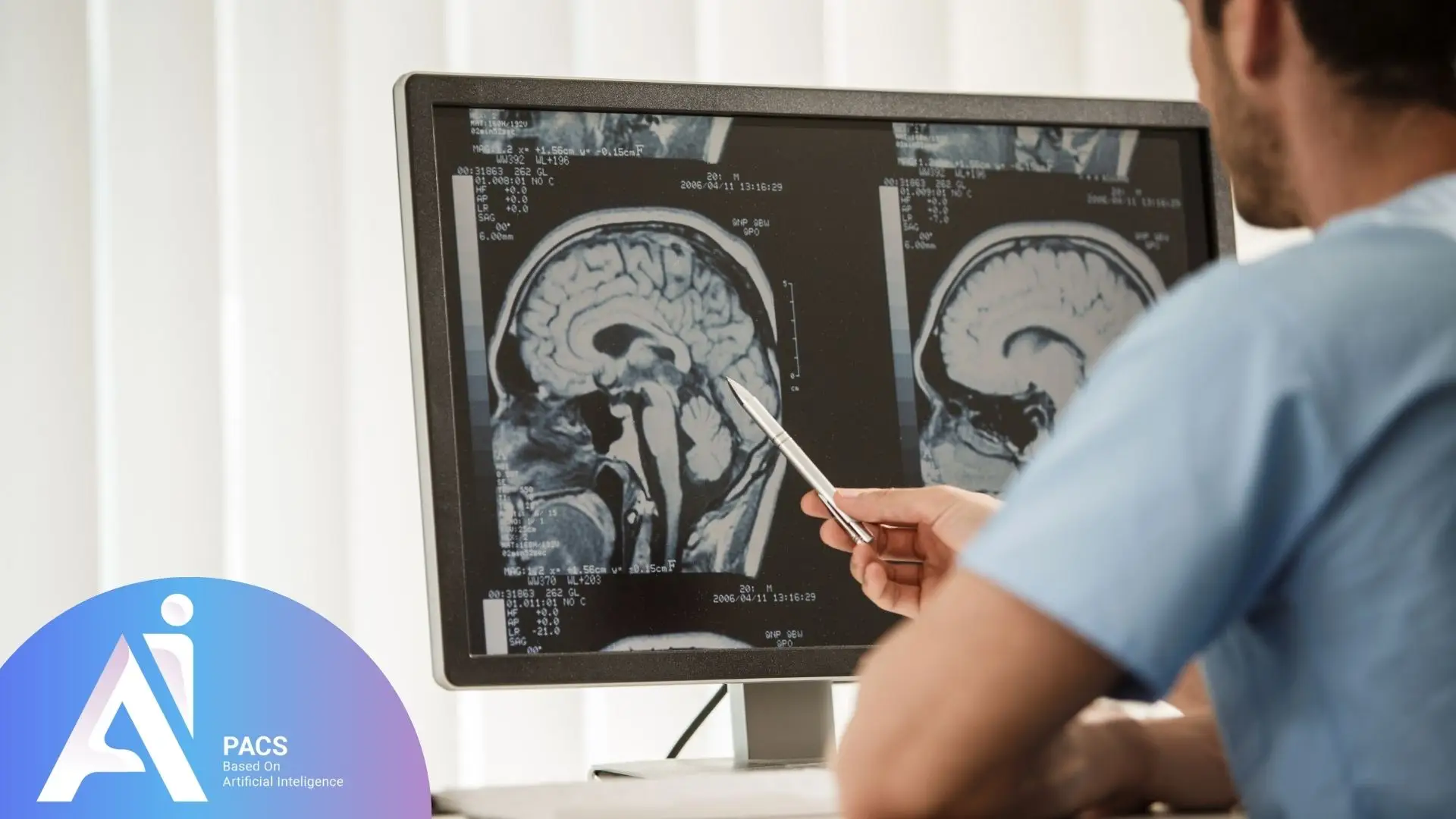

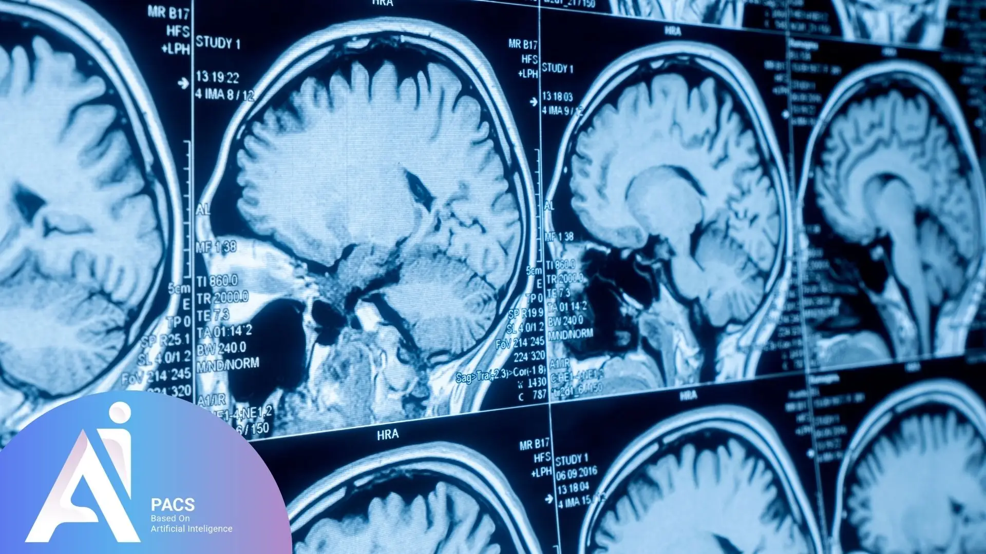
Brain MRI plays a vital role in medical diagnostics by offering clear, detailed images of brain structures, helping doctors detect and monitor various neurological conditions. As a non-invasive and highly accurate imaging technique, it has become an essential tool in modern medicine. Compared to other imaging modalities like CT scans, which are more effective in detecting acute hemorrhages and fractures, MRI excels in soft tissue contrast and provides superior visualization of brain abnormalities, making it a preferred method for diagnosing neurological conditions and tracking disease development over time. Accurate interpretation of MRI reports is crucial for diagnosing conditions such as tumors, strokes, and neurodegenerative disorders, allowing doctors to make informed treatment decisions and improve patient outcomes.
Applications of brain MRI
Detecting Brain Tumors
MRI can accurately identify tumors and abnormal masses in the brain, determining their location, size, and type.
Assessing Brain Injuries
This technique can reveal injuries caused by trauma, accidents, or other impacts, providing precise details on the location and severity of the damage.
Identifying Infections
MRI can detect brain infections such as brain abscesses, encephalitis, and meningitis, pinpointing the location and severity of the infection.
Evaluating Vascular Issues in the Brain
MRI is capable of identifying vascular problems such as aneurysms, blockages, and malformations, helping assess blood flow within the brain.
Diagnosing Neurological and Degenerative Diseases
MRI aids in diagnosing neurological conditions such as multiple sclerosis (MS), Parkinson’s disease, Alzheimer’s disease, and other degenerative brain disorders.
Examining Structural Brain Abnormalities
This technique can detect structural abnormalities like hydrocephalus (fluid accumulation in the brain), brain malformations, and congenital defects.
Analyzing Epilepsy and Other Seizure Disorders
MRI can identify brain regions responsible for seizures, aiding physicians in developing appropriate treatment plans.
Evaluating Surgical and Treatment Outcomes
After brain surgeries or other treatments, MRI helps assess outcomes and monitor the patient’s condition.
Investigating Vision and Hearing Problems
MRI can detect issues related to the optic and auditory nerves, contributing to a more accurate diagnosis of vision and hearing impairments.
It is important to note that each of these evaluations requires specific MRI sequences and techniques, making this a highly complex field. For example, the FLAIR (Fluid-Attenuated Inversion Recovery) sequence is commonly used to detect multiple sclerosis lesions, while the DWI (Diffusion-Weighted Imaging) sequence is essential for identifying acute ischemic strokes. The explanations provided here offer a general overview.


Advantages and limitations
Benefits of Brain MRI
- Produces high-resolution, detailed images of internal brain structures.
- Non-invasive and painless.
- Does not use ionizing radiation.
Disadvantages and Limitations
- Relatively high cost.
- Lengthy imaging process.
- MRI may not be suitable for individuals with certain metal implants or medical devices such as older pacemakers; however, many modern MRI-compatible implants are available, and patients should consult their doctor to determine MRI safety in their specific case.
Specialized Brain MRI Terminology
Terms Related to Lesions and Abnormalities
- Lesion: An abnormal area within brain tissue.
- Tumor: An abnormal mass that may be benign or malignant.
- Edema: Swelling due to fluid accumulation.
- Hemorrhage: Bleeding into brain tissue.
- Ischemia: Reduced blood supply to a brain region.
- Stroke: Disruption of blood flow to part of the brain.
- Infarct: Tissue death due to lack of blood supply.
- Plaque: Deposits of fat or protein in blood vessels.
Terms Related to Brain Structures
- Cortex: The outer layer of the brain responsible for advanced functions such as thinking and memory.
- White matter: Brain regions composed of nerve fibers.
- Gray matter: Brain regions containing nerve cell bodies.
- Ventricles: Fluid-filled spaces within the brain.
- Basal ganglia: Deep brain structures involved in movement control.
- Hippocampus: A structure essential for memory and learning.
- Thalamus: A central brain region that transmits sensory information to the cortex.
Final Thoughts
Interpreting brain MRI scans is a key responsibility of radiologists, ensuring precise diagnosis of brain abnormalities and disorders, which is essential for effective treatment planning. These high-resolution images provide detailed insights into brain structures, aiding in diagnosing conditions such as tumors, strokes, and neurological diseases. Accurate interpretation requires extensive expertise and experience to ensure correct diagnosis and appropriate treatment. With continuous advancements in MRI technology, its role in early diagnosis and personalized treatment plans continues to expand, ultimately leading to better patient outcomes and improved healthcare strategies.
References: