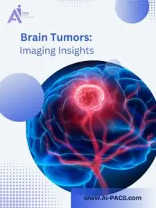


Cerebellopontine Angle Lesions: Importance of Imaging, Symptoms, and Treatment
Early recognition of lesions in the cerebellopontine (CP) angle is crucial. This area, nestled between the cerebellum and pons, houses several critical cranial nerves and vital brainstem structures. The most prevalent lesion in this area is the acoustic schwannoma (also known as a vestibular schwannoma), which primarily affects cranial nerves VII (facial) and VIII (vestibulocochlear).
I’m Dr. Vahid Alizadeh. In this article from the “When, Why, Who” series, I will explain the role of imaging in diagnosing CP angle lesions and how to manage them effectively.
Why Is Imaging Important for CP Angle Lesions?
Imaging, especially MRI with contrast, is the gold standard for detecting lesions in the CP angle. These lesions can compress multiple cranial nerves and brainstem areas, resulting in a range of symptoms.
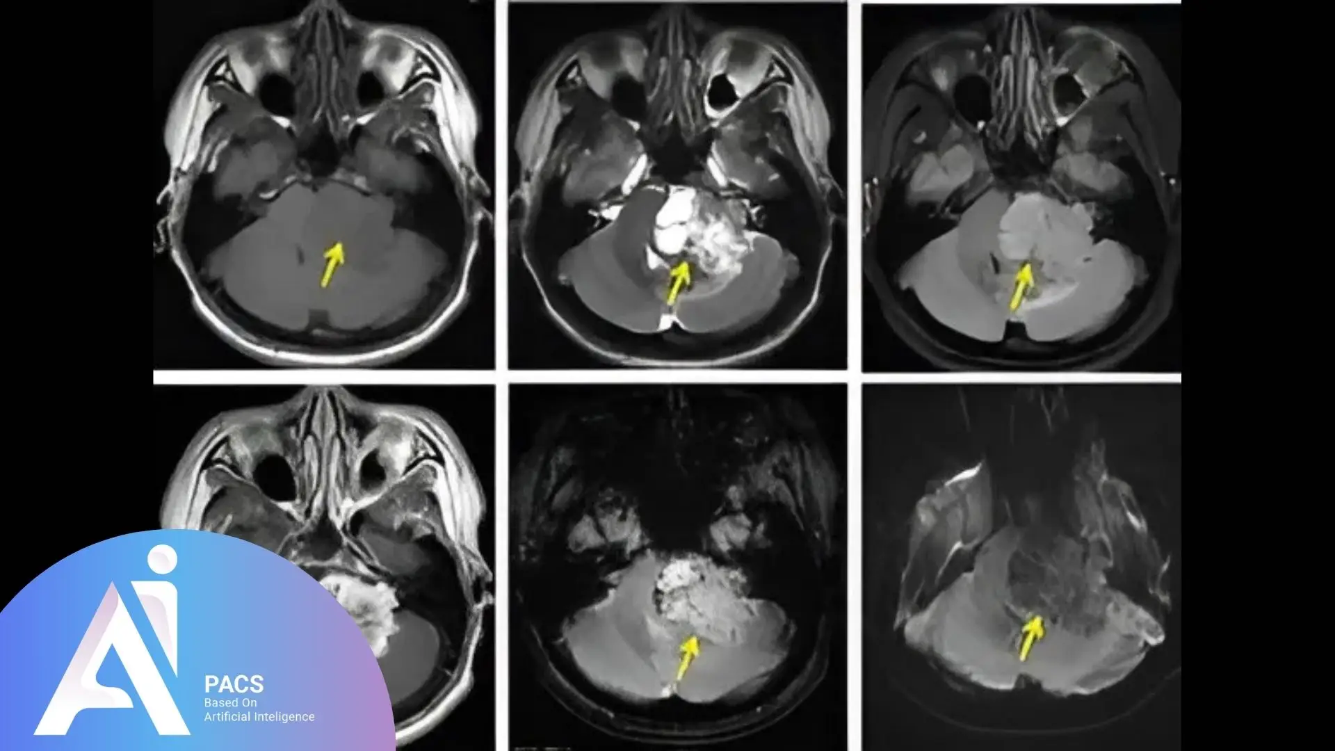
Most Common CP Angle Tumor: Acoustic Schwannoma
- Arises from the vestibular portion of the 8th cranial nerve
- Slowly growing, but may compress the 7th nerve, leading to facial weakness
- It may also press on the 5th nerve (trigeminal) or brainstem if large
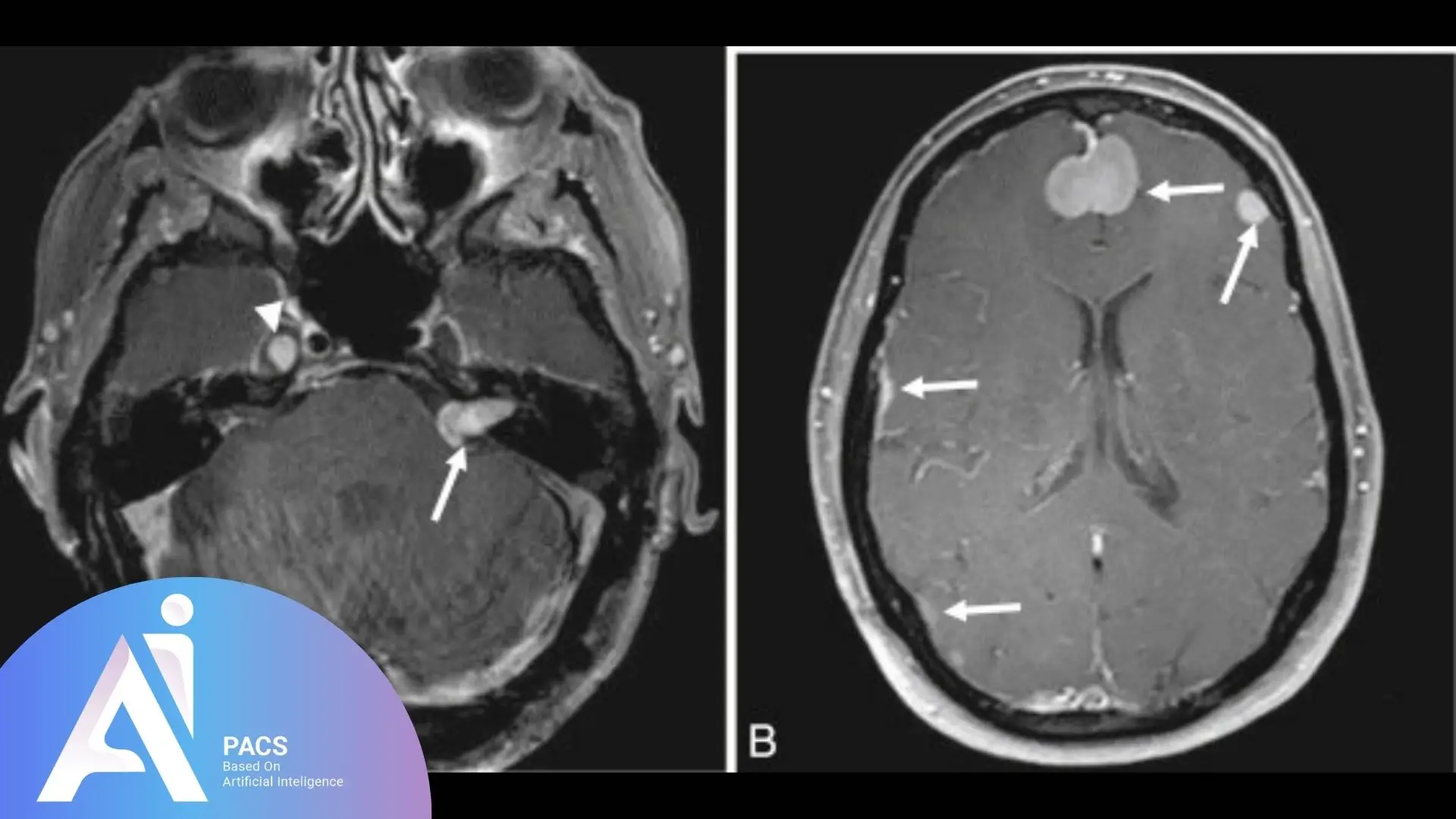
Other Common Lesions:
- Meningiomas (second most common in this area)
- Epidermoid cysts
- Rarely: metastases, arachnoid cysts, or lipomas
Importance of Cranial Nerves in the CP Angle:
- CN V (Trigeminal): Facial sensation and chewing
- CN VII (Facial): Facial expression and taste from the anterior tongue
- CN VIII (Vestibulocochlear): Hearing and balance
- Other cranial nerves passing nearby: CN IX (Glossopharyngeal), X (Vagus), XI (Accessory), and XII (Hypoglossal)
These nerves are tightly packed in this small space. Even small lesions can cause symptoms.
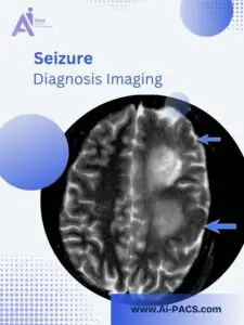
When Should Imaging Be Done for CP Angle Symptoms?
Patients may present with subtle signs that progress over time. Early imaging is a powerful tool that can prevent irreversible nerve damage, providing reassurance and confidence in the diagnostic process.
Common Symptoms That Warrant MRI:
- Unilateral hearing loss or tinnitus
- Imbalance or vertigo
- Facial numbness or twitching
- Weakness of facial muscles
- Difficulty swallowing or hoarseness (if other lower cranial nerves are involved)
MRI with contrast provides detailed views of the CP angle, showing tumor size, location, and nerve involvement.
Who Should Get Imaging and Expert Review?
Acoustic schwannomas are most common in adults aged 30–60. Although often benign, they require careful monitoring or surgery, depending on their size and symptoms.
Prevalence and Diagnosis:
- Acoustic schwannomas account for about 80% of CP angle tumors
- Incidence is roughly 1 per 100,000 people per year
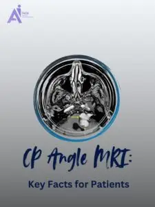
- Diagnosis is confirmed by MRI of the internal auditory canals (IAC)
Surgical and Treatment Approaches:
- Microsurgery (retro sigmoid, translabyrinthine, or middle fossa approach)
- Stereotactic radiosurgery (Gamma Knife or CyberKnife) for small tumors
- Observation for slow-growing, small tumors in older patients or asymptomatic cases
Multidisciplinary care involves collaboration among neurosurgeons, ENT specialists, and radiologists.
AI-PACS Is With You
Unclear diagnosis? Need a second opinion before surgery? AI-PACS.com offers expert reviews of MRI and CT scans, especially for brain and cranial nerve imaging.
You can upload your scan securely and receive a detailed second opinion on our online radiology report services.
Final Thoughts
Lesions in the CP angle, mainly acoustic schwannomas, can affect multiple cranial nerves and brainstem functions. Because these tumors grow slowly, symptoms may be missed early.
MRI helps detect these lesions before they cause irreversible problems. Understanding the anatomy and getting an expert imaging review is key.
Trust your instincts and trust AI-PACS to guide your next step in care.
Reference: