

Understanding CSF Leak: Key Reasons, Right Timing, and Who Needs Imaging
Cerebrospinal fluid (CSF) leaks occur when the fluid surrounding the brain and spinal cord escapes through a hole or tear in the protective membranes. One of the most common symptoms in adults is clear, watery nasal discharge, often mistaken for allergies or sinus problems.
This type of CSF rhinorrhea may occur spontaneously, after trauma, or due to tumors affecting the skull base.
I’m Dr. Vahid Alizadeh. In this article from the “When, Why, Who” series, I’ll explain how CSF leaks are diagnosed and the role imaging plays in identifying their exact cause and location.
👉 Upload Your Brain MRI for Expert Review
Why Is Imaging Important for Diagnosing a CSF Leak?
Many patients with CSF leaks experience persistent watery drainage from one side of the nose. This drainage:
- It is usually clear and thin
- This may increase with bending over or straining
- Can happen intermittently or constantly
Given that CSF leaks can mimic common nasal symptoms, it’s imperative that imaging is used to confirm the diagnosis and pinpoint the source of the leak.
Causes of CSF Leak:
- Idiopathic leaks (no apparent reason, often due to increased intracranial pressure)
- Trauma (e.g., skull base fractures, especially involving the cribriform plate or ethmoid sinuses)
- Post-surgical leaks (e.g., after sinus or brain surgery)
- Tumors that erode into the skull base or nasal cavity
When Should Imaging Be Done for Suspected CSF Leak?
If a patient presents with unexplained watery nasal discharge—especially after head trauma or surgery—imaging is necessary to empower them with a confirmed diagnosis and guide treatment.
Best Imaging Techniques for CSF Leak Detection:
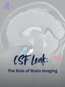
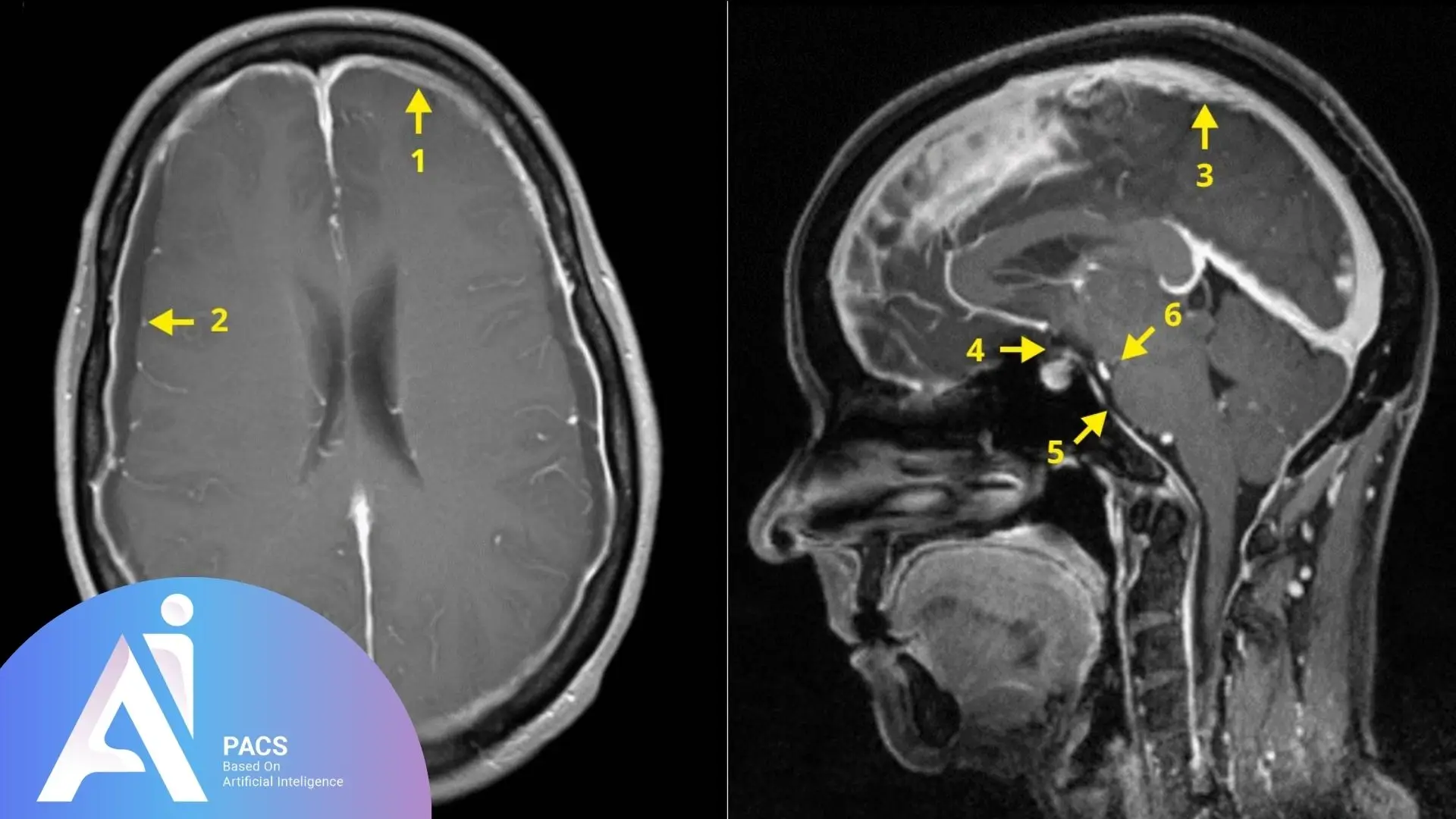
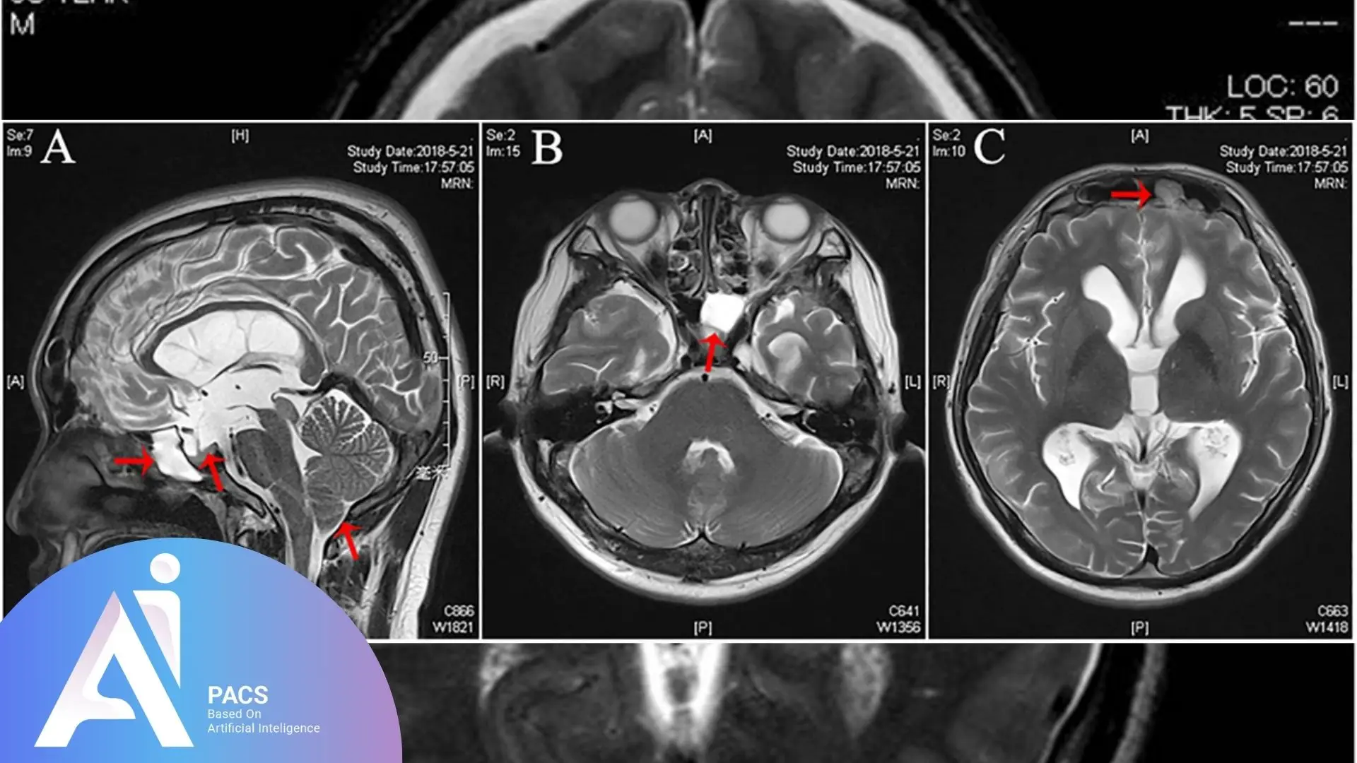
- MRI with CISS or FIESTA sequences: These sequences are highly sensitive and are used to detect fluid pathways and pinpoint leak sites, particularly in the anterior skull base.
- CT scan of the skull base: Helps identify bone defects, fractures, or erosion in areas like the ethmoid sinuses, sphenoid sinus, or cribriform plate.
How Does CT or MRI Cisternography Work?
Cisternography is a specialized imaging method used in conjunction with standard MRI or CT scans when the leak site isn’t clearly visible.
The Procedure Involves:
- Injection of contrast material into the lumbar spine (lower back) via a lumbar puncture
- The contrast then circulates with the CSF around the brain and spinal cord
- Imaging (CT or MRI) is performed to trace the flow and detect any leak points
MRI cisternography offers better contrast for soft tissue and fluid flow, while CT cisternography provides detailed images of the bone structure and any skull base defects.
This combination enables radiologists to identify the source and anatomical cause of the leak.
🖥️ If you already have your imaging and need it interpreted quickly and professionally, check out our Online Reporting Services to get expert insights without delays or extra clinic visits.
Who Needs Imaging and Expert Review?
Patients with the following symptoms or risk factors should be evaluated with brain imaging:
- Unilateral clear nasal discharge not responding to treatment
- History of head trauma or facial fracture
- Recent nasal, brain, or sinus surgery
- Signs of increased intracranial pressure
- Suspicion of skull base tumor or defect
Timely diagnosis is paramount in preventing serious complications such as meningitis or recurrent infections.
AI-PACS Is With You
If you or a loved one is experiencing symptoms of a possible CSF leak, AI-PACS.com can provide the support you need with an expert imaging review.
Our radiologists are trained to pinpoint small leaks by evaluating MRI and CT cisternography, including advanced sequences such as CISS.
Final Thoughts
CSF leaks can go unnoticed or be misdiagnosed as routine nasal issues. Clear nasal discharge may be the only sign of a serious skull base defect in adults.
Advanced imaging techniques, such as MRI CISS, CT skull base, or cisternography, are key to accurate diagnosis and successful treatment.
Trust AI-PACS to guide you to the right care and diagnosis.
Reference: