


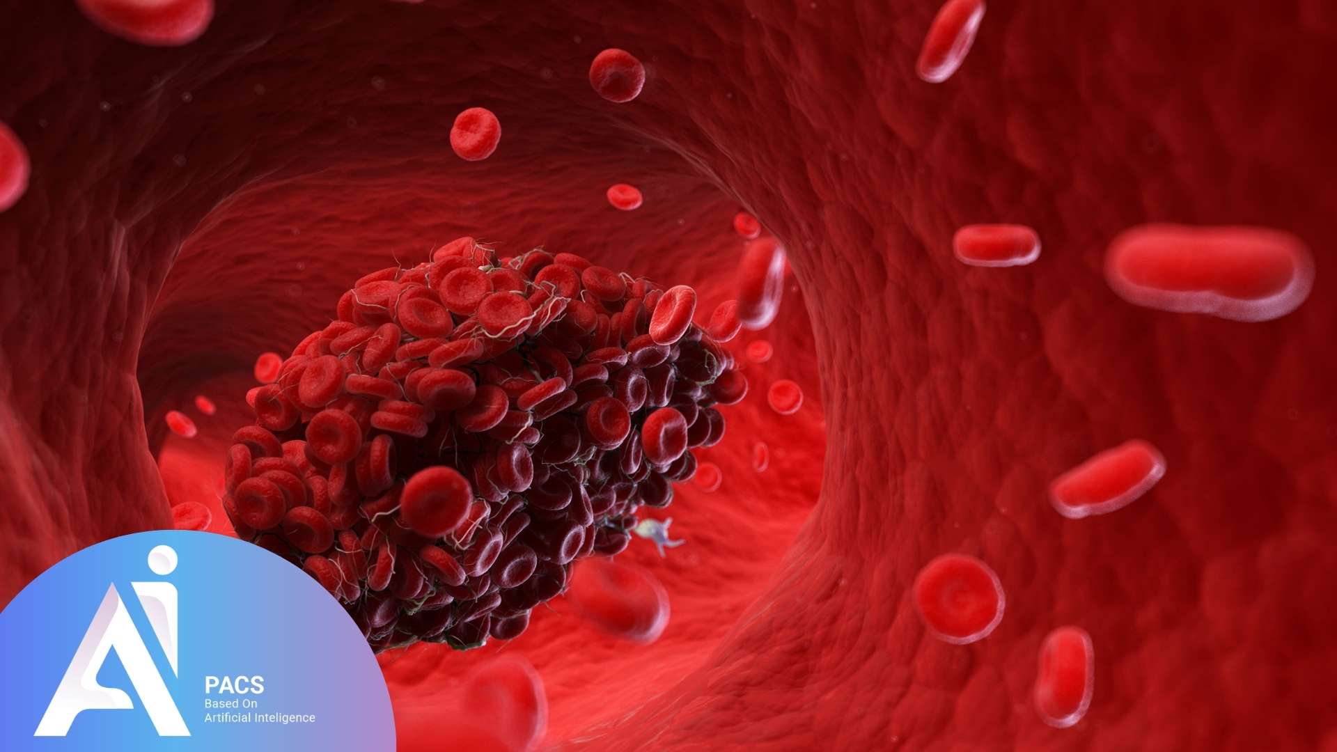
What Is a Blood Clot (Thrombosis)?
Under normal conditions, blood flows in a liquid state through the vessels. However, in certain situations—such as damage to the vessel walls, changes in blood flow, or coagulation disorders—the blood may solidify and form a clot. This clot may remain in place or travel to other parts of the body. Studies show that a significant percentage of deep vein thrombosis (DVT) cases lead to pulmonary embolism if left untreated, making early detection and intervention crucial. If it moves and becomes lodged in critical vessels, such as the arteries of the lungs or brain, it can lead to severe complications.
If you’re seeking reliable online radiology report services, AI-PACS is here to provide you with the best second opinion on your medical images, delivered by our team of expert radiologists.
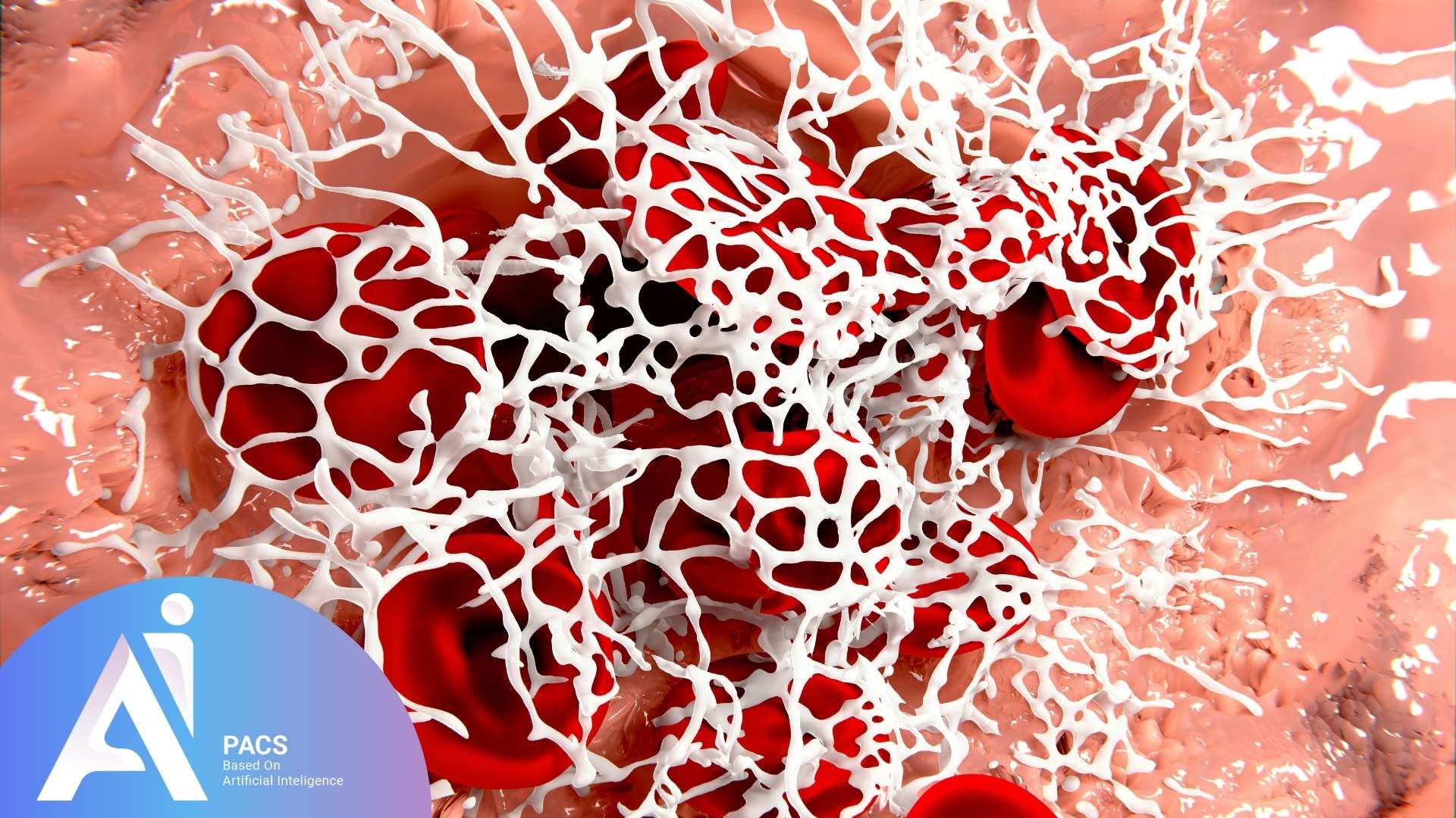
What Causes Blood Clots?
Blood clotting is a natural and essential process that stops bleeding after an injury. But sometimes clots form unnecessarily inside vessels. This happens due to a combination of factors known as Virchow’s triad:
- Injury to blood vessel walls (due to surgery, trauma, or inflammation)
NOTE: Motor vehicle accidents or falls requiring trauma imaging - Changes in normal blood flow (slow or turbulent flow from immobility or varicose veins)
- Hypercoagulability (when your blood is more prone to clot, which can be genetic or caused by medications and diseases)
Together, these factors increase the risk of clot formation inside your veins or arteries.
Types of Thrombosis
Understanding the different types of thrombosis is crucial, as they require distinct diagnostic approaches and treatment strategies. Proper identification can help determine the urgency of intervention and guide appropriate medical care.
Blood clots are generally categorized into two main types:
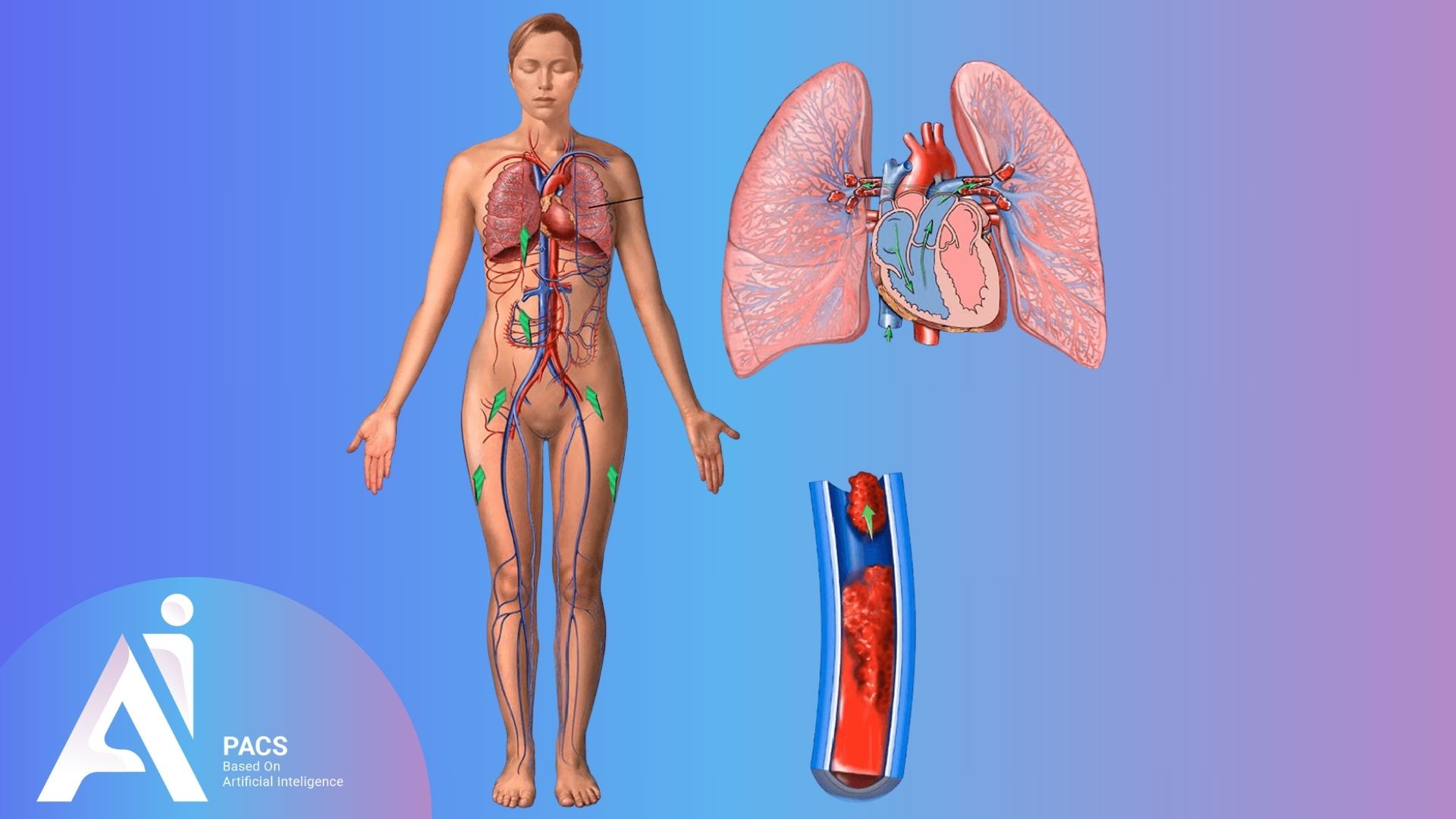
Venous Thrombosis
This type of clot forms in the veins, which are responsible for returning blood to the heart. The most common type is deep vein thrombosis (DVT), usually occurring in the legs. If a clot dislodges and travels to the lungs, it can result in a pulmonary embolism (PE)—a life-threatening condition.
Arterial Thrombosis
Arterial clots form in arteries, which carry oxygen-rich blood to the body. These clots can cause severe conditions such as heart attacks or strokes. This type of clot often develops quickly, leading to immediate symptoms and requiring urgent medical intervention.
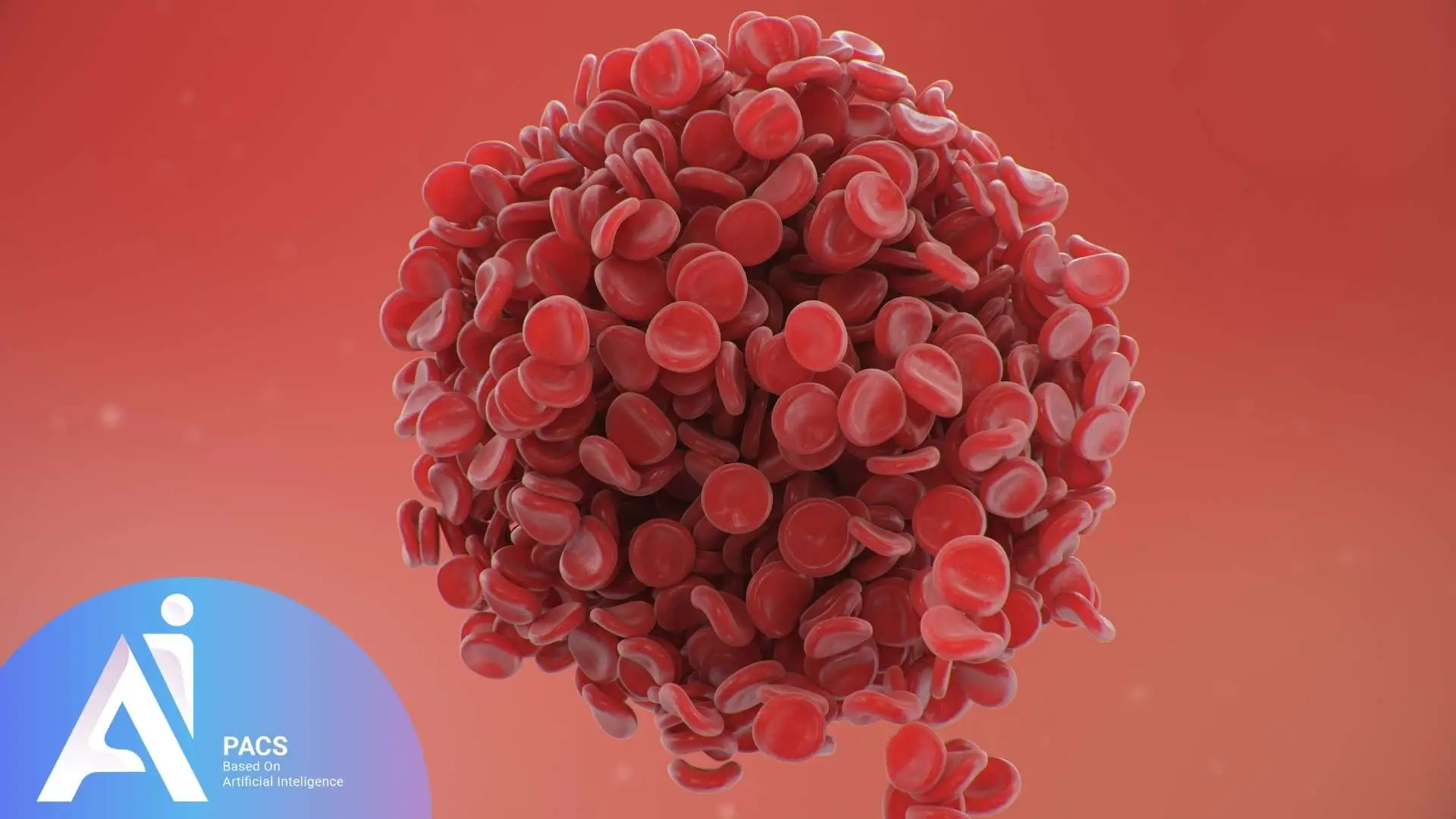
How Dangerous Are Blood Clots?
Blood clots can vary from harmless to life-threatening based on their size and location. A small clot in a superficial vein may cause mild discomfort, while a clot obstructing blood flow to the lungs or brain can lead to organ damage, permanent disability, or death. Early recognition and treatment can prevent serious outcomes. That’s why recognizing the signs and seeking medical help promptly is crucial.
For immediate expert review of your vascular imaging, our emergency CT consultation service provides rapid interpretation when time is critical.
Risk Factors for Blood Clots
Several factors can increase the likelihood of blood clot formation, including:
- Prolonged immobility – Sitting or lying down for extended periods, such as during long flights or hospitalization, increases the risk.
- Surgery or injury – Major surgical procedures or injuries can contribute to clot formation.
- Pregnancy – Increased pressure on veins and hormonal changes during pregnancy elevate the risk.
- Obesity – Excess weight can put additional pressure on blood vessels.
- Smoking – Smoking damages blood vessels, making clot formation more likely.
- Chronic diseases – Conditions such as cancer, diabetes, or heart disease can heighten the risk.
- Family history – A genetic predisposition to blood clots may increase susceptibility.
- Certain medications – Oral contraceptives and hormone replacement therapy can contribute to clotting.
Get Fast and Accurate Analysis of Your Medical Imaging
How Does a Blood Clot Present Itself?
Symptoms vary depending on the clot’s location. While some individuals may experience no symptoms, common signs include:
Deep Vein Thrombosis (DVT) – Legs
- Swelling in one leg (typically unilateral)
- Pain or tenderness, especially when standing or walking
- Redness or discoloration of the skin (bruising)
- A sensation of warmth in the affected area
Lungs (Pulmonary Embolism – PE)
- Sudden shortness of breath
- Chest pain, particularly when taking deep breaths
- Coughing, sometimes with blood
- Rapid or irregular heartbeat
Stroke
- Sudden weakness or numbness on one side of the body
- Difficulty speaking or understanding speech
- Vision problems or loss of balance
- Severe and sudden headache
Imaging Techniques for Blood Clot Diagnosis
Medical imaging plays a crucial role in diagnosing blood clots, allowing healthcare providers to determine their location, size, and severity, and to administer the most appropriate treatment. The choice of imaging technique depends on the clot’s location and the patient’s condition.
Ultrasound (Doppler Sonography)
This is the first-line imaging method for evaluating DVT in the legs. It uses sound waves to create images of blood flow within the vessels. Doppler ultrasound helps detect changes in circulation and identify blockages.
Advantages: Fast, safe, painless, and does not require contrast dye.
CT Scan (Computed Tomography)
CT scans, particularly CT angiography, are used to diagnose clots in the abdomen, lungs (PE), and brain.
- How it works: A contrast dye is injected into the bloodstream, producing high-resolution images of the blood vessels.
- Advantages: High accuracy in detecting clots, especially in critical areas, and provides rapid results.
MRI (Magnetic Resonance Imaging)
MRI is often used for assessing clots in complex areas such as the brain, spine, or deeper veins.
How it works: MRI uses magnetic fields and radio waves to generate high-resolution images.
Advantages: No radiation exposure and provides highly detailed images.
Angiography
This method is often preferred when other imaging techniques do not provide sufficient clarity or when real-time visualization of blood flow is necessary for accurate diagnosis and treatment planning. It is used for precise identification of clots in the arteries of the heart or brain.
How it works: A contrast dye is injected into the arteries, allowing real-time visualization of blood flow.
Advantages: The best method for diagnosing clots in coronary and cerebral arteries, especially in patients with suspected stroke or heart attack.
Each imaging method hasspecific advantages for different types of clots. Expert vascular imaging interpretation ensures youreceive accurate diagnosis and appropriate urgency assessment for your specific situation.
Additional Tests
Alongside imaging, doctors may conduct blood tests to measure D-dimer levels. Elevated D-dimer levels can indicate the presence of a clot somewhere in the body.
Treatment Options for Blood Clots
Treatment for blood clots depends on their location and severity. Common approaches include:
Anticoagulant medications (blood thinners): Prevent further clot formation and reduce risk of complications.
Thrombolytic therapy (clot-busting drugs): Used in emergencies to dissolve dangerous clots.
Surgical procedures: In severe cases, procedures such as thrombectomy (surgical removal of the clot) or insertion of a vena cava filter may be necessary.
Supportive care: Compression stockings, leg elevation, and lifestyle changes.
If you’ve received imaging for suspected blood clots, professional radiology consultation ensures accurate interpretation of your vascular studies.
Key Strategies for Preventing Blood Clots
Preventing blood clots involves managing risk factors and adopting a healthy lifestyle. Here are some essential prevention tips:
- Engage in regular physical activity, especially if you sit for extended periods.
- Stay hydrated to prevent blood thickening.
- Avoid smoking.
- Use compression stockings if recommended by a doctor.
- Take prescribed anticoagulants if you are at high risk.
- Move around frequently during long trips (e.g., standing and walking in an airplane or train).
Final Thoughts
Blood clots are a serious medical condition that can occur at any age and pose significant health risks. Awareness of symptoms and early detection through medical imaging can facilitate timely diagnosis and treatment. Advances in imaging technology enable doctors to identify clots with greater accuracy and determine the best course of action. Treatment options, including anticoagulants and surgical interventions, can effectively manage the condition. If you experience any suspicious symptoms, do not ignore them—seek medical attention immediately.
Blood clot diagnosis through imaging requires expert interpretation to distinguish between different types of clots and assess their clinical significance. If you’ve undergone imaging for suspected blood clots, professional radiological consultation can help you understand your results and their implications for your treatment and long-term health.
References: