


What is MR Angiography?
Magnetic Resonance Angiography (MRA) is a non-invasive diagnostic imaging technique that allows for the examination of blood vessels and blood flow. It is used to detect narrowing, blockages, and other vascular abnormalities. MRA utilizes a strong magnetic field, radio waves, and a computer system to generate high-resolution 2D and 3D images, helping physicians diagnose various vascular diseases. This method does not use ionizing radiation, making it safer than some other imaging techniques. In some cases, a contrast agent may be injected before imaging to enhance the visibility of blood vessels.
Applications of MRA and Its Importance
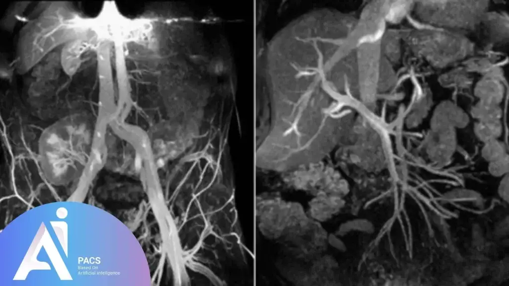
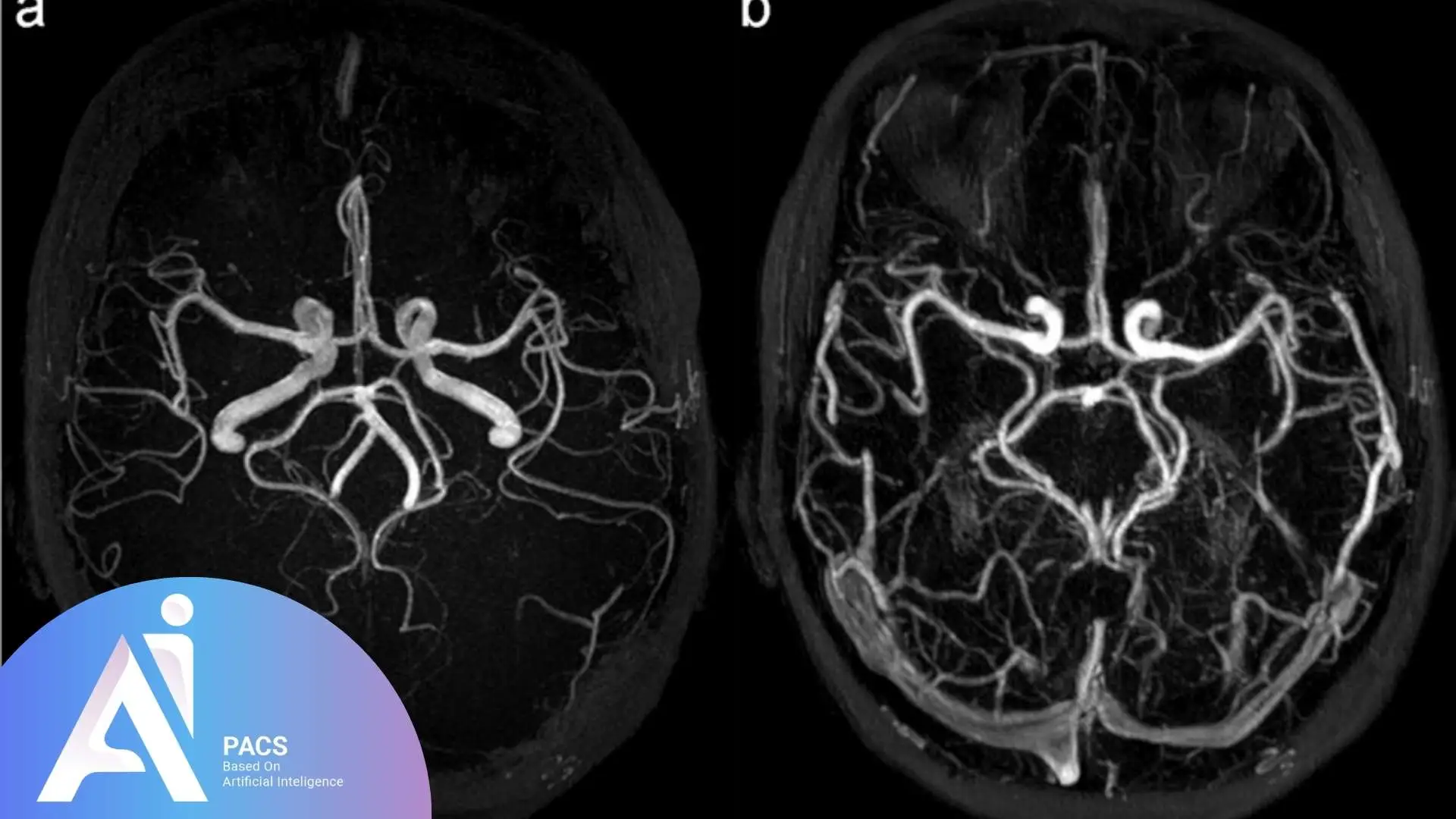
As mentioned earlier, MRA is used to assess blood vessels. Its applications include evaluating the blood vessels in the brain, neck, heart, chest, abdomen (such as the kidneys and liver), pelvis, arms, and legs. It is also used to detect abnormalities like aortic aneurysms, vascular stenosis, blockages, and diseases such as atherosclerosis (plaque buildup) in the carotid artery of the neck, which can reduce blood flow to the brain and increase the risk of stroke.
With MRA, physicians can obtain detailed images of the structure and function of the body’s blood vessels, which is highly beneficial for both diagnosis and treatment. For example, in stroke patients, MRA can help identify vascular blockages early, enabling timely intervention and reducing the risk of severe complications.
Who Should Undergo an MRA?
MRA is recommended for individuals with vascular diseases or those at risk of developing them. Candidates for MRA include:
- Individuals experience symptoms such as chest pain, heart discomfort, shortness of breath, muscle weakness or drooping, or changes in skin color, which may be related to cardiovascular diseases.
- Individuals with a family history of cardiovascular diseases, including diabetes, high blood pressure, high cholesterol (hyperlipidemia), and smoking.
- Patients with inconclusive vascular imaging results who require more detailed imaging.
- Individuals experiencing vascular changes in specific areas such as the brain, neck, heart, or legs.
Who Should Avoid MRA?
Before undergoing MRA, it is essential to inform your physician about any existing medical conditions, recent surgeries, allergies, or pregnancy. Although magnetic fields are generally safe, they may interfere with certain medical devices. Most orthopedic implants pose no risk, but individuals with pacemakers or metal implants should inform the technician before the procedure.
Inform your healthcare provider before the test if you have:
- Brain aneurysm clips
- Artificial heart valve
- Heart defibrillator or pacemaker
- Inner ear (cochlear) implants
- Insulin pump or chemotherapy port
- Intrauterine device (IUD)
- Kidney disease or dialysis (you may not be able to receive contrast)
- Neurostimulator
- Recently placed artificial joints
- Vascular stent
- Worked with sheet metal in the past (you may need tests to check for tiny metal pieces in your eyes)
Advantages and Disadvantages of MRA
Advantages
- Non-invasive and safe: Unlike conventional angiography, MRA does not require catheter insertion into blood vessels.
- High-resolution imaging: Provides detailed and precise images of blood vessels.
- Superior vascular diagnostics: Offers better accuracy than some other imaging techniques.
- Enhanced imaging with contrast: Some scans may require contrast agents to improve image quality and diagnostic accuracy.
Disadvantages
- Higher cost: MRA is generally more expensive than other imaging methods.
- Limitations for certain patients: Individuals with pacemakers or metal implants may not be eligible for this procedure.
- Immobility requirement: Patients must remain still during the scan, which can be challenging for some.
- Potential contrast agent reactions: Patients with allergies may experience adverse reactions to the contrast agent if required.
How MRA is Performed
The MRA procedure typically follows these steps:
- Preparation: Patients should avoid wearing metal objects, such as jewelry, watches, or glasses, and will be required to wear a disposable gown. Certain types of metal can cause blurry images.
- Positioning: The patient lies still on the examination table to ensure high-quality images.
- Contrast Injection (if needed): If a contrast agent is required, the physician should be informed of any allergies before administration. Some examinations need a special contrast dye. Usually, this dye is injected into a vein in your hand or forearm before the procedure. The contrast helps the radiologist get a clearer view of specific areas.
- Imaging Process: The MRI machine uses a magnetic field and radio waves to capture images without exposing the patient to radiation. The scanner may produce loud noises, but headphones can help reduce discomfort.
- Completion: Once the scan is completed, the images will be analyzed by a radiologist for interpretation.
Note: Patients may need to fast for 4 to 6 hours before the test, depending on the physician’s instructions.
Comparison of MRA with Other Imaging Techniques
MRA vs. CT Scan
Magnetic Resonance Angiography (MRA) and Computed Tomography (CT) scans are both imaging methods used to look inside the body, but they have some important differences:
- Radiation Exposure: MRA utilizes magnetic fields and radio waves, thereby avoiding exposure to ionizing radiation in patients. CT scans, on the other hand, rely on X-rays, which involve radiation that can increase health risks over time.
- Image Quality: MRA provides highly detailed 3D images of blood vessels, enabling doctors to view complex vascular structures with greater clarity and precision. CT scans can also produce 3D images, but MRA often offers better resolution for soft tissues and blood vessels.
- Diagnostic Accuracy: For certain vascular conditions, such as stenosis (narrowing of blood vessels) or aneurysms (vessel bulging), MRA tends to be more precise in detecting abnormalities compared to CT.
MRA vs. Ultrasound
Ultrasound is a widely used and safe imaging technique that utilizes sound waves to visualize internal structures, particularly blood flow. However, when compared with MRA:
- Image Detail and Resolution: MRA produces higher-resolution images, offering more detailed views of the blood vessels’ anatomy and function. Ultrasound images are typically less detailed and can be limited by the patient’s body type or the operator’s skill.
- 3D Imaging: MRA can generate three-dimensional images, allowing better visualization of vascular networks from multiple angles. Ultrasound generally provides two-dimensional images and limited 3D capabilities.
- Availability and Limitations: Ultrasound machines are more widely available and less costly, but may not always provide the necessary clarity for complex vascular assessments. MRA, while more detailed, may not be available everywhere and is more expensive.
MRA vs. MRI
Both MRA and MRI use magnetic fields and radio waves, but their focus and applications differ:
- MRI (Magnetic Resonance Imaging): Primarily used to capture detailed images of soft tissues such as the brain, muscles, organs, and tumors. MRI helps diagnose a wide range of conditions beyond the vascular system, including neurological disorders, injuries, and certain types of cancer.
- MRA (Magnetic Resonance Angiography): A specialized type of MRI focused on imaging blood vessels, particularly arteries. It is commonly used to detect vascular diseases such as blockages, narrowing (stenosis), or aneurysms, providing a clear map of blood flow and vessel health.
MRA vs. MR Venography (MRV)
Both MRA and MRV are magnetic resonance techniques, but target different parts of the circulatory system:
- MRA: Concentrates on imaging arteries, the vessels that carry oxygen-rich blood from the heart to the rest of the body. It helps identify arterial diseases such as atherosclerosis, stenosis, or aneurysms.
- MRV (Magnetic Resonance Venography): Specifically designed to visualize veins, which return blood to the heart. MRV is useful for diagnosing venous conditions, including deep vein thrombosis (DVT), venous thrombosis, and venous insufficiency.

Post-Test Care
After undergoing MRA, certain precautions may be necessary:
- Resting Period: Although no special recovery is required, some individuals may need a short period of rest, as advised by their physician.
- Monitoring for Allergic Reactions: If a contrast agent was used, symptoms such as swelling, itching, shortness of breath, or muscle weakness should be reported to a doctor immediately. Patients should drink plenty of fluids to help flush the contrast dye from the body.
- Following Medical Instructions: Patients should adhere to any dietary or medication restrictions prescribed by their physician.
Interpretation of MRA Results
A radiologist interprets MRA results to determine whether the findings are normal or indicate vascular abnormalities. If any issues are detected, additional tests such as CT scans or echocardiography may be required to confirm the diagnosis and guide treatment planning.
If you’re seeking reliable online radiology report services, AI-PACS is here to provide you with the best second opinion on your medical images, delivered by our team of expert radiologists.
Final Thoughts
MRA is a non-invasive diagnostic imaging technique used to detect vascular diseases, including heart and brain vessel disorders. It provides high-quality, detailed images of blood vessel structures and functions. Since MRA does not involve radiation exposure, it is considered a safe and effective diagnostic tool. As research advances, new developments in MRA, such as faster scanning times and improved contrast agents, are making the procedure even more efficient. If you suspect vascular issues, consulting a physician for further evaluation and timely intervention is advisable.
References:
radiologyinfo.org
hopkinsmedicine.org
National Institute of Health