


What is MR Venography?
Magnetic Resonance Venography (MRV) is an advanced, specialized, non-invasive, and painless imaging technique used to evaluate blood flow in veins and detect blood clots. This technology enables physicians to obtain a more precise view of the body’s venous structures and circulation.
MRV safely produces high-resolution images of internal venous structures using strong magnetic fields and radio waves, avoiding ionizing radiation. However, it has limitations, including longer scan times compared to CT venography and higher costs, making accessibility a consideration for some patients. This method aids in identifying and assessing venous obstructions, narrowing, blood clots, and other circulatory issues. Due to its high accuracy and interpretability, MRV is a valuable tool in diagnosing and monitoring conditions such as venous thrombosis, venous stenosis, and other vascular abnormalities. It also facilitates treatment planning by providing three-dimensional imaging of venous anatomy, detecting irregular or reversed blood flow, and assessing venous blockages, including heart vein occlusions.
Why is MR Venography Performed?
Diagnosis of Venous Disorders
MR Venography is primarily used for assessing veins and diagnosing venous conditions, including blood clots, narrowing (stenosis), deep vein thrombosis (DVT), or vein occlusions.
Preoperative Planning for Venous Surgery
MRV assists physicians in accurately evaluating venous structures and pathways, aiding in surgical planning and therapeutic decision-making.
Assessing Treatment Effectiveness
By comparing pre- and post-treatment images, MRV can reveal changes in venous structure and function, allowing physicians to evaluate the effectiveness of interventions such as thrombectomy, stent placement, or anticoagulant therapy for venous disorders.
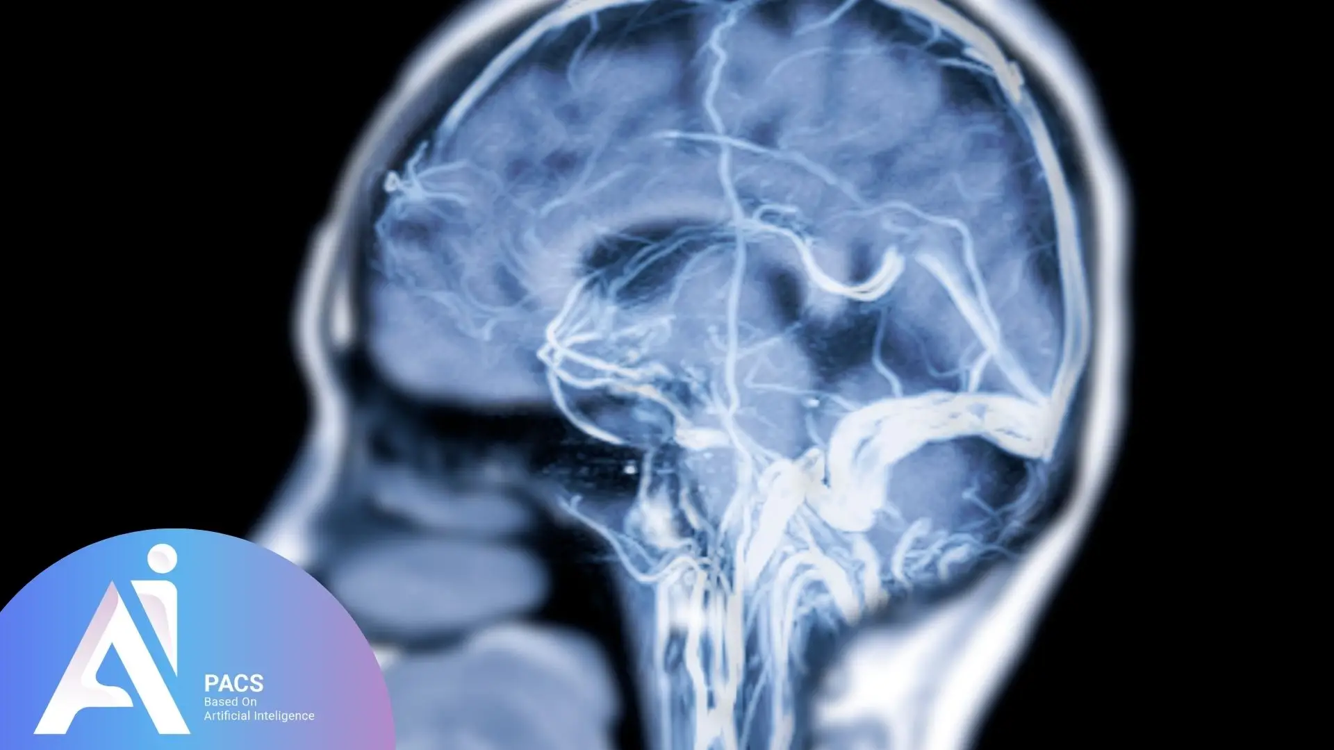
Brain MR Venography
In MRV, a contrast agent (free of ionizing radiation) is often injected into the body to enhance the image clarity of the veins. This technique allows for detailed visualization of venous structures without the use of X-rays. Brain MRV is particularly useful for diagnosing conditions such as cerebral venous thrombosis (CVT), venous structural abnormalities, intracranial hypertension, and cerebral blood flow disorders.
Other Common Indications for MRV
- Diagnosis of congenital venous anomalies
- Identifying suitable veins for bypass graft surgery
- Determining the cause of leg pain
- Locating the origin of a blood clot
- Evaluating deep vein valve function
- Investigating blood circulation issues
- Diagnosing normal pressure hydrocephalus (NPH)
- Identifying intracranial pressure problems
- Detecting deep vein thrombosis (DVT)
Preparation Before MRV
Patients should inform their physician or imaging center staff about any medications they are taking, as well as any known allergies, particularly to contrast agents.
Before entering the MRI room, patients must remove any electronic or metallic objects such as watches, mobile phones, jewelry, or other accessories, as these items can interfere with the magnetic field and affect image quality. If a contrast agent is used, patients may be required to fast for approximately four hours before the test. Additionally, pre-procedural testing may be necessary to confirm that the patient can safely receive the contrast agent. Providing accurate medical history and treatment records can help the physician or MRI technician interpret the results more precisely.

How is MRV Performed?
Patient Preparation
Before the procedure, patients may be asked to change into a hospital gown to prevent any interference from clothing with metallic components. They should also inform their physician of any medical implants or conditions that might affect the scan. If contrast is required, patients may need to undergo kidney function tests to ensure safe administration of the contrast agent.
Procedure Execution
The patient lies on a table that slides into the MRI scanner. In cases requiring higher image quality, an intravenous contrast agent is administered. During the scan, the patient should remain still to prevent motion artifacts and noise interference on the images.
The MRI machine generates strong magnetic fields and radio waves to capture venous images. These signals are processed into three-dimensional images, which the physician then uses to analyze venous structures. Upon completion, the images are provided to the physician for further diagnosis and evaluation.
Potential Risks and Side Effects of MRV
MRV is a safe and non-invasive procedure with minimal side effects. However, some patients may experience minor adverse reactions:
Contrast Agent Reactions
Some individuals may experience mild allergic reactions to contrast agents, though these are generally rare and manageable. Patients with kidney disease who receive contrast agents are at a higher risk of severe reactions, which can affect tissues throughout the body, including the skin, joints, liver, and lungs.

Who Should Avoid MR Venography?
Certain groups should avoid it or take precautions, including:
- Patients with implanted medical devices, such as pacemakers, cochlear implants, or metal clips, should be cautious because the magnetic field can interfere with these devices.
- Individuals with severe claustrophobia or anxiety may have difficulty staying calm during the scan without sedation.
- People with impaired kidney function, as the contrast dye used in MRV, can increase the risk of kidney-related complications.
- Pregnant women are usually advised to avoid MRV unless it is necessary due to potential dangers from contrast agents.
Before scheduling an MRV, it’s important to discuss your medical history with your doctor to ensure the procedure is safe for you.
Advantages of MRV
MRV offers several significant advantages compared to other imaging techniques like CT venography:
No Ionizing Radiation: Unlike X-ray or CT scans, MRV uses magnetic fields and radio waves, making it safer, especially for patients requiring repeated imaging.
High-Quality Imaging: The high contrast of MRV images allows for better differentiation of veins from surrounding tissues, improving diagnostic accuracy.
Contrast-Free Imaging Option: In some cases, MRV can be performed without a contrast agent, which is beneficial for patients with allergies or those unable to tolerate contrast materials.
Three-Dimensional Visualization: MRV provides 3D imaging, allowing physicians to assess venous structures from multiple angles and gain a better understanding of the patient’s vascular condition.
No Need for Anesthesia: MRV typically does not require anesthesia, making it safer and more accessible for a wide range of patients, including children. However, its longer scan duration may be challenging for patients who struggle to remain still for extended periods.
Broad Diagnostic Applications: MRV is effective in diagnosing a wide range of venous conditions, including deep vein thrombosis, nutcracker syndrome, venous malformations, and other vascular disorders.
Interpreting MRV Results
MRV images are analyzed by radiologists specializing in vascular imaging, who undergo extensive training in interpreting venous structures, identifying abnormalities, and assessing blood flow patterns to provide accurate diagnoses. The interpretation process involves several critical steps requiring expertise and experience:
1. Assessing Image Quality
The radiologist evaluates the clarity of MRV images to ensure they are suitable for interpretation. This includes checking for sharpness, motion artifacts, or any distortions that may affect accuracy.
2. Identifying Venous Structures
The radiologist examines the size, shape, and presence of abnormalities such as narrowing, blockages, or aneurysms in the veins.
3. Evaluating Blood Flow
MRV allows visualization of venous blood flow. The radiologist looks for signs of reduced or disrupted flow, which may indicate clot formation or other vascular issues.
4. Diagnosing Abnormalities
MRV images are used to detect congenital or acquired venous abnormalities, including conditions affecting major veins such as cerebral sinuses, limb veins, and the inferior vena cava.
5. Reporting Findings
After image analysis, the radiologist compiles a detailed report describing the findings and, if necessary, recommends further diagnostic tests or treatments. If you’re seeking reliable online radiology report services, AI-PACS is here to provide you with the best second opinion on your medical images, delivered by our team of expert radiologists.
Final Thoughts
While MRV has numerous advantages, the choice of imaging modality should be based on the patient’s specific needs, medical condition, and availability of imaging equipment. Compared to CT venography, MRV avoids ionizing radiation and provides superior soft tissue contrast, but CT may be preferred for rapid emergency assessment. Unlike ultrasound, which is widely available and cost-effective, MRV offers more detailed three-dimensional imaging but is less accessible and more expensive. By carefully analyzing MRV images and findings, physicians and radiologists can significantly enhance the diagnosis and management of venous disorders, ensuring optimal patient care.
References: