
Online Second Opinion for Breast Cancer by Expert Radiologists
Get an expert second opinion on your breast MRI, mammogram, or ultrasound—secure, accurate, and fully online.
Upload Your Breast Imaging
Meet the Experts Consulting on Your Breast Cancer Case
Your images are independently reviewed by three radiologists, who discuss the findings. The lead radiologist prepares the written opinion for you.
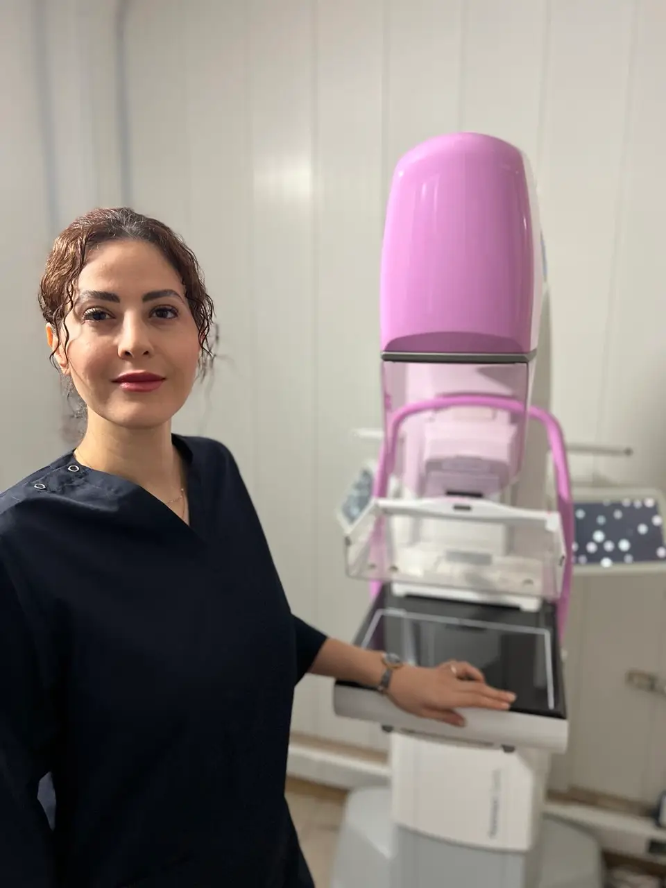
Dr. Bahare Mobasheri
Radiologist
Fellowship‑trained in breast imaging with clinical focus on
diagnostic and screening mammography.
MC-NO: 133284

Dr. Vahid Alizadeh
RadiologistExtensive experience interpreting complex mammography cases and second‑opinion reviews. Member of the European Society of Radiology (ESR) | ESR ID: 160956
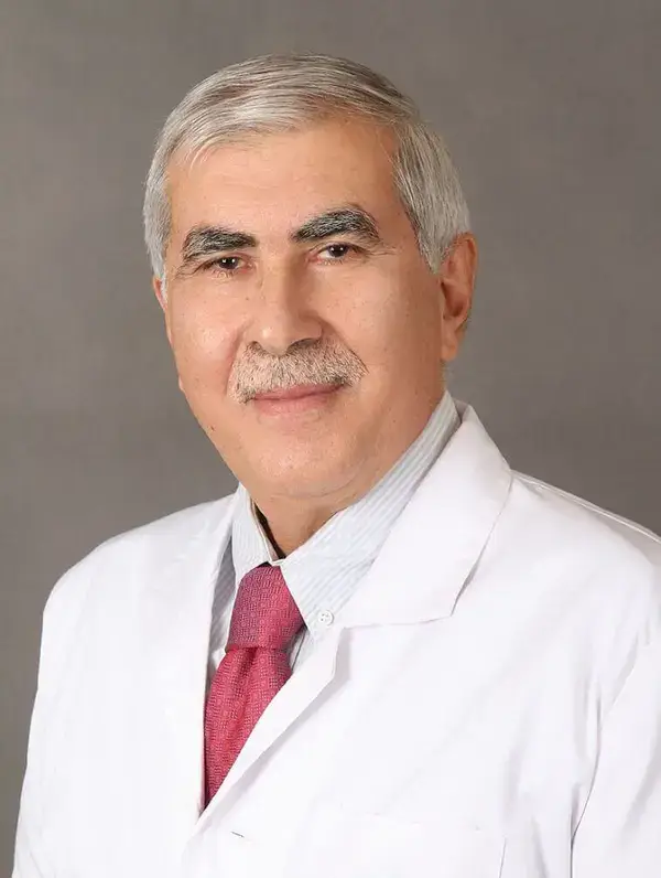
Dr. Reza Alizadeh
Radiologist
Focused expertise in correlating mammography findings with
ultrasound and breast MRI when needed.
40 Years of Diagnostic Excellence
What We Review for Your Breast Cancer Second Opinion?
Share the relevant breast imaging and clinical information so our team can confirm what can be reviewed. Diagnostic interpretation requires complete DICOM images; supporting reports or screenshots do not replace them.
-
Breast Imaging Studies
Mammogram, breast MRI, or ultrasound images, including screening and diagnostic views when available. For a dedicated review of ultrasound studies, see our breast ultrasound second opinion service. - Original report for later clarification
We usually do not ask for the original report before the independent image review. If questions remain after delivery, we may ask you to share it so the team can explain differences in wording or interpretation. -
Prior Breast Imaging (If Available)
Previous mammograms, MRIs, or ultrasounds to help identify changes over time and provide context. -
Additional Supporting Information
Any related reports or clinical details you choose to share to support a more comprehensive review.
Bringing your breast imaging findings together
“My mammogram, ultrasound and breast MRI were performed at different centers. How do their findings relate to each other?”
A breast imaging second opinion can bring the relevant examinations you provide into one written review. Tell us which finding or recommendation you want clarified and share your complete imaging studies in DICOM format, with their dates and relevant clinical information.
The review explains how the findings relate, whether the available images support the same interpretation, and what remains uncertain. Mammography, ultrasound and MRI show different aspects of breast tissue, so differences between their reports do not automatically mean that one report is wrong.
Our service focuses on radiology interpretation. Your treating team considers the imaging opinion alongside examination findings and any biopsy results when making care decisions.
Comparing breast imaging during follow-up or treatment
“I have several examinations from different dates. What has changed, and what can be compared reliably?”
For an Advanced Consultation, our team can review the relevant examinations and medical records you provide to summarize imaging changes over time. The team confirms the review scope after checking the material.
To help the team assess a comparison, include:
- Earlier and current images: complete available studies, with the date and imaging center for each examination.
- Clinical context: relevant surgery or treatment dates and supporting records, when they relate to your question.
- Your main questions: the finding, apparent change or difference between examinations you want explained.
Differences in technique, incomplete studies or missing earlier images can limit comparison. The written summary identifies these limits and helps you discuss the imaging findings with your treating team; imaging alone does not establish treatment success or determine a cancer treatment plan.
What you receive from a breast imaging second opinion
You receive a written radiology opinion explaining the relevant findings, the assessment and important limitations of the supplied images. You can ask questions about the report after delivery.
Explore the fictional breast MRI example below to see how an imaging opinion addresses a patient's question. This example contains one MRI examination; it does not demonstrate a completed mammogram–ultrasound–MRI comparison. Your own report reflects your images, clinical information and agreed review scope.
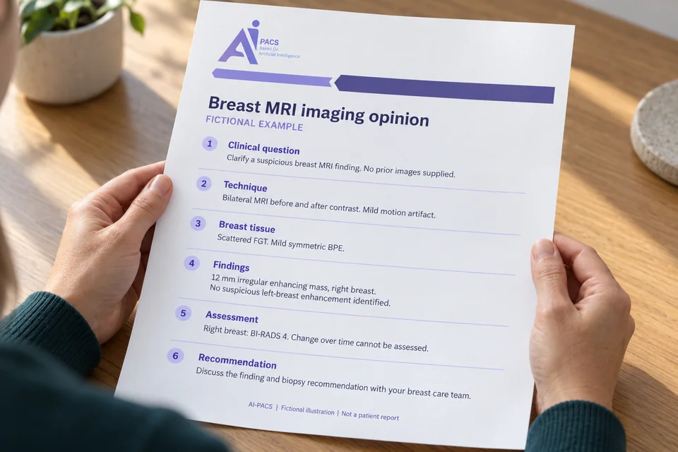
01 Clinical question and comparison
Clinical question and comparison
In this fictional example: Independent review requested to clarify a suspicious finding on breast MRI. No prior mammogram, ultrasound or MRI images supplied for comparison.
Your questions give the review a focus. The report identifies which studies were actually available. This example cannot establish change over time or agreement with another imaging method.
02 Technique and image quality
Technique and image quality
In this fictional example: Bilateral breast MRI with T2-weighted images and pre- and post-contrast T1-weighted sequences. Mild motion artifact; the described mass remains assessable.
Technique describes how the examination was acquired, including whether contrast images are available. Motion, uneven fat suppression or missing sequences can limit interpretation; the relevant limitation should be explained.
03 Breast tissue: FGT and BPE
Breast tissue: FGT and BPE
In this fictional example: Scattered fibroglandular tissue (FGT). Mild, symmetric background parenchymal enhancement (BPE).
FGT describes the amount of fibrous and glandular breast tissue seen on MRI. BPE describes contrast enhancement of background breast tissue. These describe the tissue background and are separate from the suspicious finding below.
04 Relevant abnormal and normal findings
Relevant abnormal and normal findings
In this fictional example: A 12 mm irregular mass with heterogeneous enhancement is seen in the upper outer right breast. No additional suspicious enhancement is identified in the left breast on this examination.
Findings record the location, size and visible features of an abnormality, together with relevant observations without a corresponding abnormality. The left-breast statement is limited to this MRI; it is not a guarantee that every possible condition has been excluded.
05 Assessment and impression
Assessment and impression
In this fictional example: Suspicious enhancing mass in the right breast: BI-RADS 4. No prior images are available to assess interval change.
The impression brings the observations into an imaging assessment. BI-RADS 4 describes a suspicious finding for which biopsy is recommended; it does not establish a tissue diagnosis of cancer. A second opinion may support the original assessment.
06 Recommendations and your questions
Recommendations and your questions
In this fictional example: Discuss the suspicious finding and biopsy recommendation with the breast care team. Further imaging correlation may help determine an appropriate biopsy approach.
Recommendations connect the assessment to the next clinical discussion. After delivery you can ask about the wording and remaining uncertainty. If you need clarification of differences from the original report, the team may then ask you to share it. An imaging review should not delay recommended assessment or biopsy.
Want an independent review of your breast imaging? Share your complete DICOM studies, relevant clinical information and the questions you want clarified. Include earlier examinations with their dates if your question concerns changes over time.
Request a breast imaging second opinion