

Understanding Mammogram Results
A mammogram is a low-dose X-ray test used to look for breast changes before they can be felt. It is one of the most important screening tools for breast cancer, especially in women at average or increased risk.
But like every medical test, mammography is not perfect. Sometimes it suggests something abnormal that later turns out to be harmless. In other cases, it may miss a cancer that is actually present. These two situations are called false positives and false negatives.
Mammography has limits, and some findings need more evaluation before a final answer is available. If you already have a report and want an independent review of the images, see our Mammogram Second Opinion service. A second read may agree or disagree with the first; it cannot guarantee that every cancer will be detected.
What Is a False Positive Mammogram?
A false positive mammogram happens when the screening test shows an area that looks suspicious, but follow-up testing shows there is no cancer.
Why does this happen?
Common reasons include:
- overlapping normal breast tissue
- breast cysts
- benign calcifications
- fibroadenomas
- technical image limitations
- changes from prior surgery or inflammation
Sometimes the radiologist may recommend:
- extra mammogram views
- breast ultrasound
- breast MRI in selected cases
- short-interval follow-up imaging
- biopsy if a finding still looks concerning
Does a false positive mean the radiologist made a mistake?
Not necessarily. In screening mammography, the goal is to detect even subtle abnormalities early. That means some women will be recalled for additional testing even though the finding is ultimately benign. This is a known and expected part of screening.
How common are false positives?
False positives are not rare, especially in first-time mammograms or in women with dense breasts. The chance of recall depends on age, breast density, prior comparisons, and the screening method used.
A recall often follows an unexpected result on a routine scan — see what an abnormal mammogram can mean and what happens next.
False-positive results can lead to additional appointments, costs, and worry. Complete the recommended assessment before treating a recalled finding as benign; a callback is not yet a confirmed false positive.
What Is a False Negative Mammogram?
A false negative mammogram happens when the mammogram appears normal, but breast cancer is actually present.
Why can mammograms miss cancer?
A mammogram may miss a cancer because:
- the tumor is very small
- the breast tissue is dense
- the cancer is in a difficult location
- the lesion blends into surrounding tissue
- the image quality is limited
Are false negatives dangerous?
A false negative can delay diagnosis, which is why ongoing screening and attention to breast symptoms matter. Still, mammograms detect many cancers early, and the overall benefit of screening is substantial.
Can symptoms appear even after a normal mammogram?
Yes. A normal mammogram does not rule out every breast problem. If you notice a lump, skin thickening, nipple discharge, persistent pain in one area, or nipple changes, you should seek medical evaluation even if your recent mammogram was normal.
How Accurate Are Mammograms?
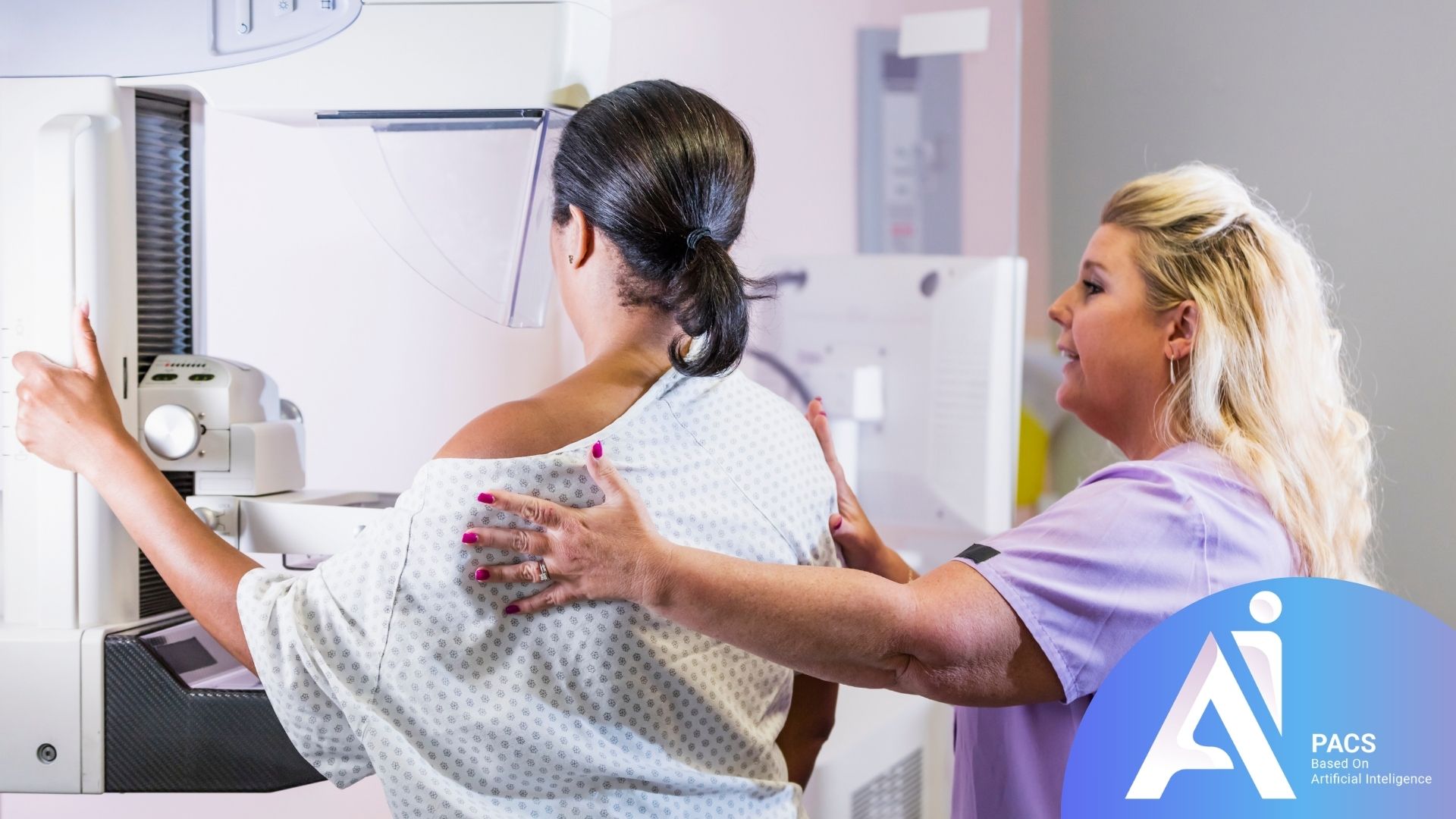
There is no single “accuracy percentage” that applies to every mammogram. Sensitivity, specificity, and the chance that a recalled finding is cancer describe different things. Results also depend on the population, technique, and whether the examination is screening or diagnostic.
Sensitivity and specificity
- Sensitivity means how well a test finds disease when it is present.
- Specificity means how well a test correctly identifies people without disease.
Screening mammography has good performance overall, but accuracy varies depending on:
- breast density
- age
- hormonal status
- whether the study is digital mammography or tomosynthesis
- the radiologist’s experience
- whether previous images are available for comparison
Dense breasts and mammogram accuracy
Dense breast tissue appears white on a mammogram, and many breast cancers also appear white. That can make detection harder. Women with dense breasts may have a higher chance of both false positives and false negatives.
Discuss supplemental screening with your clinician based on overall risk and breast density. Ultrasound or MRI may be appropriate in selected situations; neither is automatically required after every normal mammogram. Tomosynthesis is a mammographic technique, not a guarantee against missed cancer.
What Happens If You Are Called Back After a Mammogram?
Being called back does not mean you have cancer.
A callback usually means the radiologist wants a better look at an area that was not fully clear on the screening images.
Common next steps include
- repeat mammogram with magnification or spot compression
- ultrasound
- comparison with prior images
- biopsy if the area still looks suspicious
Many callbacks end with reassurance and no cancer diagnosis.
How should you think about a callback?
Think of it as a “please check more carefully” result, not a “you have cancer” result.
Scientific Explanation in Simple Terms
Breast imaging works by identifying patterns in tissue density, calcifications, masses, and distortion. Radiologists compare the image appearance with known benign and malignant patterns.
Mammogram interpretation often uses the BI-RADS system:
- 0: incomplete; additional imaging and/or prior-image comparison needed
- 1: negative
- 2: benign finding
- 3: probably benign, short-term follow-up
- 4: suspicious; biopsy generally recommended
- 5: highly suggestive of malignancy; biopsy generally recommended
- 6: biopsy-proven cancer
A screening mammogram is designed to detect possible abnormalities early. Because it prioritizes sensitivity, it may flag findings that later prove harmless. That is the tradeoff that helps screening find cancers sooner.
Read for more information: What Does BIRADS Mean in Mammography?
Practical Advice for Patients
Before your mammogram
- Bring prior breast imaging if possible.
- Tell the clinic about breast surgery, implants, or prior biopsies.
- Schedule the test when your breasts are less tender if that helps you tolerate compression better.
After your mammogram
- Read the report carefully with your clinician.
- Do not panic if you are called back.
- Ask what the BI-RADS category means.
- Ask whether you need short-term follow-up or a biopsy.
If you have dense breasts or higher risk
- Ask whether supplemental screening is appropriate.
- Discuss personal and family history with your doctor.
- Do not skip follow-up appointments.
If your mammogram is normal but you have symptoms
- Do not ignore the symptom.
- Ask for a clinical breast exam.
- Ask whether ultrasound or diagnostic imaging is needed.
What Research Shows—and Its Limits
Research helps describe screening performance across groups. It cannot determine whether one person’s mammogram is correct or replace follow-up of a specific symptom or finding.
1) False positives can affect future screening behavior
A 2024 study reported in the Annals of Internal Medicine and highlighted by the National Cancer Institute found that women who experienced false-positive mammograms were somewhat less likely to return for future routine screening. This is important because regular screening helps detect breast cancer earlier.
Source: National Cancer Institute, 2024.
2) Tomosynthesis improves some screening measures
Durand and colleagues’ 2021 Radiology study found better sensitivity and specificity with tomosynthesis than digital mammography. False-negative rates showed a downward trend, but the difference was not statistically significant. The study does not establish that 3D mammography prevents missed cancers or improves survival.
Source: Durand et al., Radiology, 2021.
3) Dense breast tissue remains a major challenge
Dense tissue can obscure cancers on a mammogram. Decisions about supplemental imaging should consider the person’s overall risk as well as density, including the possibility of additional false-positive findings.
Source: RadiologyInfo.org: Dense Breasts.
4) Interval cancers remain an important research topic
An interval cancer is diagnosed between scheduled screening examinations. Some were not visible or were missed at the earlier examination; others develop or grow afterward. An interval diagnosis alone does not prove that a radiologist overlooked a visible cancer.
Source: National Cancer Institute: Breast Cancer Screening.
When Should You Contact a Doctor?
You should contact a healthcare provider if:
- your mammogram report is unclear
- you were told to return for more imaging
- you feel a new lump
- you notice nipple discharge, skin dimpling, or breast asymmetry
- breast pain is persistent and localized
- you have a strong family history of breast cancer and have questions about screening
Early communication helps avoid delay and reduces anxiety.
Conclusion
Yes, a mammogram can sometimes be wrong, but the more accurate way to say it is that it can produce false positives or false negatives. This is a normal limitation of medical screening tests, not a reason to distrust mammography.
The good news is that mammograms remain one of the most effective tools for early breast cancer detection. If you are called back for more tests, it usually means the radiologist wants to be careful—not that cancer is already confirmed.
Scientific References Used
- National Cancer Institute: Mammograms
- National Cancer Institute: False-positive results and future screening (2024)
- Durand et al. False-Negative Rates of Breast Cancer Screening with and without Digital Breast Tomosynthesis. Radiology. 2021;298:296–305.
- RadiologyInfo.org (ACR/RSNA): Mammography
- RadiologyInfo.org (ACR/RSNA): Dense Breasts
- National Cancer Institute: Breast Cancer Screening