
What Does “Suspicious Finding” on a Mammogram Mean?
When you receive a mammogram report mentioning a “suspicious finding,” it can understandably cause worry. However, this term does not mean that cancer has been diagnosed. In a final breast imaging assessment, “suspicious” indicates concern that may require a biopsy. Read the BI-RADS category and recommendation: a callback for an incomplete examination is different from a final suspicious assessment.
Mammography is used both for screening and to investigate symptoms or an abnormal screening result. An incomplete screening result (BI-RADS 0) may need additional views, ultrasound, or comparison with prior images. It should not automatically be described as BI-RADS 4 or cancer.
How Radiologists Classify Mammogram Findings
Radiologists typically use the BI-RADS (Breast Imaging Reporting and Data System) classification developed by the American College of Radiology. This standardized system helps describe imaging findings and guide follow‑up care.
Key BI‑RADS Categories Related to Suspicious Findings
- BI-RADS 0: Incomplete — additional imaging and/or comparison with prior images is needed.
- BI-RADS 1: Negative. BI-RADS 2: A benign finding.
- BI-RADS 3: Probably benign (no more than 2% likelihood of cancer); follow-up imaging is recommended.
- BI-RADS 4: Suspicious abnormality; a biopsy is generally recommended.
- BI-RADS 5: Highly suggestive of malignancy; a biopsy is strongly recommended.
BI-RADS 4 covers a wide range of suspicion; 4A, 4B, and 4C are not interchangeable. The assessment is not a tissue diagnosis. BI-RADS 6 is used when a biopsy has already confirmed cancer.
Read for more information: What Does BIRADS Mean in Mammography?
What Types of Changes Can Appear Suspicious on a Mammogram?
Radiologists analyze several imaging features that may indicate an abnormality. Suspicious findings can include:
Breast Masses
A mass is a dense area that stands out from surrounding breast tissue. Certain characteristics—such as irregular shape or poorly defined edges—may make it appear suspicious.
Microcalcifications
These are tiny calcium deposits that appear as white specks on mammograms. Most calcifications are benign, but certain patterns or clusters may require closer examination.
A single prominent white area raises different questions than scattered specks — see what a large bright white spot on a mammogram can mean.
Architectural Distortion
This refers to abnormal pulling or distortion of the breast tissue structure without a visible mass. It can sometimes indicate early cancer or scar tissue.
Asymmetry
If one area of the breast appears denser or different from the corresponding area in the other breast, it may prompt additional imaging.
Why Additional Imaging Is Often Recommended
If a suspicious finding appears on a screening mammogram, your doctor may recommend a diagnostic mammogram. This exam uses additional views and magnified images to better evaluate the area of concern.
Other imaging methods may include:
- Breast ultrasound: helps determine whether a mass is solid or fluid-filled.
- Breast MRI: may be used for selected clinical questions, high-risk screening, or evaluating known cancer. It is not routinely needed for every callback and does not replace mammography or a recommended biopsy.
These additional tests help radiologists better characterize the abnormality and decide whether a biopsy is necessary.
When Is a Biopsy Needed?
For a final BI-RADS 4 or 5 assessment, tissue sampling is generally recommended. The care team chooses the biopsy method using the imaging appearance and clinical findings. A biopsy removes a small tissue sample for laboratory analysis; not every uncertain finding requires one.
Common biopsy techniques include:
- Core needle biopsy (most common)
- Stereotactic biopsy guided by mammography
- Ultrasound‑guided biopsy
- MRI‑guided biopsy
Most breast biopsies are minimally invasive and performed with local anesthesia. Importantly, many biopsies reveal benign conditions such as cysts, fibroadenomas, or benign calcifications.
How Often Are Suspicious Findings Actually Cancer?
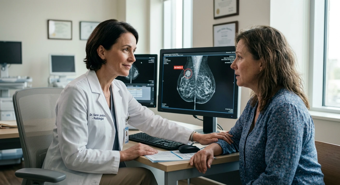
Many patients assume that “suspicious” automatically means cancer, but this is not the case.
There is no single cancer percentage for every suspicious mammogram. BI-RADS 4 spans more than 2% to less than 95% likelihood of malignancy and is often subdivided:
- 4A: more than 2% and up to 10%.
- 4B: more than 10% and up to 50%.
- 4C: more than 50% and less than 95%.
BI-RADS 5 indicates at least 95% likelihood, but tissue diagnosis is still needed. These category ranges are not a personalized diagnosis or a reason to skip the recommended biopsy. Ask your care team which category applies to your report.
What Patients Should Do After Receiving This Result
Receiving a report mentioning a suspicious finding can be stressful, but there are practical steps you can take:
- Schedule recommended follow‑up imaging promptly.
- Discuss the BI‑RADS category with your physician or radiologist.
- Ask whether additional imaging or biopsy is necessary.
- Keep copies of your imaging reports for future comparison.
It is important to remember that early evaluation provides clarity and allows doctors to rule out serious conditions or begin treatment early if necessary.
Practical Breast Health Tips for Patients
In addition to routine mammography, maintaining awareness of breast health can help detect changes earlier.
- Follow screening guidelines based on your age and risk factors.
- Perform breast self-awareness rather than rigid monthly exams—be familiar with how your breasts normally feel.
- Report any new lumps, nipple discharge, skin changes, or persistent pain to your healthcare provider.
- Maintain a healthy lifestyle, including regular physical activity and limiting alcohol consumption.
When Should You Seek a Second Opinion?
If you feel uncertain about your results or recommended next steps, seeking a second opinion from another radiologist or breast imaging specialist can be helpful.
Second opinions are particularly valuable when:
- A biopsy is recommended.
- Imaging findings are unclear.
- You have a strong family history of breast cancer.
An independent image review can assess the reported finding and explain whether the reviewing radiologist agrees with the imaging recommendation. See our Mammogram Second Opinion service for the review process, or request a review of your mammogram. Provide the original DICOM images, report, and relevant prior studies. A second opinion may agree with the original report and should not delay recommended imaging or biopsy.
Conclusion
A suspicious mammogram assessment is not a confirmed cancer diagnosis, but it deserves the follow-up recommended in the report. The BI-RADS category, imaging features, and clinical findings determine whether additional imaging or biopsy is appropriate.
If you receive this result, the most important step is to complete the recommended follow‑up testing. Early investigation provides reassurance when findings are benign and allows prompt treatment if necessary.