
What Is a Mammogram vs 3D Mammography (Tomosynthesis)?
A mammogram is a specialized, low-dose X-ray of the breast used to screen for and diagnose breast cancer. The conventional form, called 2D digital mammography, takes two flat pictures of each breast—one from top to bottom and one from side to side. For decades, this has been the gold-standard tool for finding cancers before they can be felt.
3D mammography, known medically as digital breast tomosynthesis (DBT), is a newer evolution of the same technology. Instead of capturing only two flat images, the machine sweeps in a small arc and takes many low-dose X-rays from different angles. A computer then reconstructs these into a series of thin “slices,” much like the pages of a book, allowing the radiologist to scroll through the breast layer by layer.
The simplest way to picture the distinction: a standard mammogram is like looking at a single photograph of a crowd, where people can hide behind one another. Tomosynthesis is like walking through that crowd, so overlapping figures separate out. This is the core idea behind the mammogram vs 3D mammography (tomosynthesis) comparison.
How Does Each Exam Work?
The Standard 2D Mammogram
During a 2D mammogram, your breast is gently positioned on a flat plate and compressed by a second paddle for a few seconds. The X-ray machine then takes one image, and the process repeats for a second angle and the other breast. The compression spreads the tissue so that abnormalities show up more clearly and so less radiation is needed. The whole appointment usually takes about 10 to 20 minutes.
The 3D Mammogram (Tomosynthesis)
A 3D exam feels almost identical from the patient’s side—the same positioning and the same brief compression. The difference happens inside the machine. The X-ray tube moves in an arc above the breast, capturing multiple low-dose images across that path. Software stacks these into dozens of thin cross-sections. Most modern systems acquire both a 3D dataset and a “synthetic” 2D image from the same scan, so a separate 2D exposure is often no longer required. This keeps the total radiation low while giving the radiologist far more detail to review.
Key Differences at a Glance
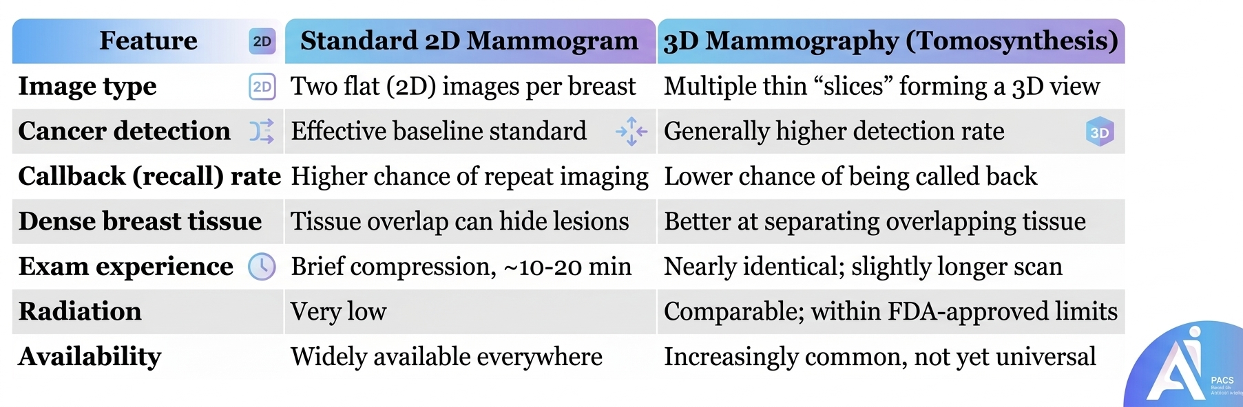
The table below summarizes the practical differences in the mammogram vs 3D mammography (tomosynthesis) discussion.
Who Benefits Most from 3D Mammography?
While tomosynthesis offers advantages for most women, certain groups tend to benefit the most.
Women with dense breast tissue often gain the clearest advantage. Dense tissue appears white on a mammogram—and so does cancer—so a tumor can blend into the background like a snowball in a snowstorm. Because tomosynthesis separates overlapping layers, it can reveal lesions that a flat image might hide. Many regions now require that women be notified of their breast density, so this is an increasingly relevant point.
Read for more information: Can Dense Breast Tissue Affect Mammogram Results?
Women who have experienced frequent callbacks in the past may also appreciate 3D screening, since it tends to reduce the number of false alarms that lead to repeat imaging and anxiety. That said, a standard 2D mammogram remains a thoroughly effective and validated screening tool, and not every woman needs the 3D version. Your decision should reflect your breast density, personal and family history, and what equipment is available locally. This is exactly the kind of nuance worth discussing with your physician.
Diagnosis & What to Expect During Your Exam
Preparing for either exam is straightforward. On the day of your appointment, avoid using deodorant, antiperspirant, powders, or lotions on your chest and underarms, because metallic particles in these products can appear as spots on the images. Wearing a two-piece outfit makes undressing from the waist up easier.
During the test, a technologist will position each breast on the imaging plate and apply compression for a few seconds. Compression can feel tight, but it should not be painful; if you have tender breasts, scheduling the exam in the week after your period may help. The actual imaging takes only moments.
A radiologist—a physician trained to interpret medical images—reviews your results and reports findings using a standardized scale called BI-RADS. If something needs a closer look, you may be asked to return for additional imaging such as a diagnostic mammogram or ultrasound. Being recalled does not mean you have cancer; most callbacks turn out to be benign.
Accuracy, Recall Rates, and Radiation
A common question in the mammogram vs 3D mammography (tomosynthesis) conversation is whether the newer technology is truly more accurate. The evidence increasingly says yes, with meaningful but modest gains.
In terms of cancer detection, 3D mammography consistently finds slightly more cancers per thousand women screened than 2D alone. Just as importantly, it lowers the recall rate, meaning fewer women are called back unnecessarily for additional pictures—a benefit that reduces both cost and worry.
On radiation, patients often assume that “more images” means “more radiation.” In practice, the dose from a modern 3D exam is very low and falls within the safety limits set by regulators. When a synthetic 2D image is generated from the 3D scan, the combined dose stays comparable to a traditional two-view mammogram. For nearly all women, the proven benefit of early detection far outweighs this small radiation exposure.
Practical Tips for Patients
- Ask what your facility offers. Not every clinic has 3D equipment. If you have dense breasts or a strong family history, ask whether tomosynthesis is available to you.
- Know your breast density. If your report or notification mentions dense tissue, discuss whether 3D mammography or additional imaging (such as ultrasound or MRI) is appropriate.
- Check coverage in advance. Insurance coverage for 3D mammography has expanded but can still vary; a quick call to your provider avoids surprises.
- Bring prior images. Comparing this year’s exam with previous ones helps the radiologist spot subtle changes and reduces unnecessary callbacks.
- Stay on schedule. Whichever method you choose, regular screening matters far more than the specific technology. Consistency is what saves lives.
- Don’t panic over a callback. Being asked to return for more imaging is common and usually ends with reassuring news.
If findings are complex, using AI PACs Mammogram Second Opinion Online Service can provide additional reassurance and expert review.
Latest Scientific Findings
Recent research strengthens the case for tomosynthesis while confirming that standard mammography remains valuable.
According to a 2024 study published in Radiology (RSNA), a ten-year analysis found that screening with digital breast tomosynthesis increased cancer detection rates and significantly reduced the rate of advanced cancers compared to conventional 2D digital mammography. The same research reported that the recall rate for DBT was 7.2%, significantly lower than the 10.6% for digital mammography. For patients, this suggests cancers may be caught earlier while fewer women face the stress of being called back. Radiological Society of North AmericaScienceDaily
A large 2023 cohort study from the Breast Cancer Surveillance Consortium, published in Radiology, examined repeat screening rounds and found that recall rates remained lower for DBT than for digital mammography, while cancer detection rates were modestly higher. This matters because it shows the benefit is not limited to a woman’s first 3D exam—it persists over years of routine screening. PubMed Central
A 2022 systematic review and meta-analysis in European Radiology concluded that tomosynthesis combined with synthesized 2D mammography is associated with higher cancer detection and lower recalls compared to digital mammography alone. Pooling many studies adds confidence that these advantages are real and reproducible. arxiv
Finally, the U.S. Preventive Services Task Force, in its April 2024 update, recommended that all women be screened every other year starting at age 40 and continuing through age 74. Notably, the Task Force also stated that the evidence was insufficient to assess the benefits and harms of DBT as a primary screening method, meaning experts still consider both 2D and 3D mammography acceptable screening choices. US Preventive Services TaskforceUS Preventive Services Taskforce
Conclusion
When it comes to mammogram vs 3D mammography (tomosynthesis), the most important message is reassuring: both are effective, safe tools for finding breast cancer early. The traditional 2D mammogram remains a trusted standard, while 3D tomosynthesis offers added detail that can detect more cancers and reduce unnecessary callbacks—benefits that are especially meaningful for women with dense breast tissue.
There is no single “right” answer for everyone. The best choice depends on your breast density, your personal risk, and what your local facility offers. What matters most is that you screen regularly and stay engaged with your care.
If you have questions about your results or want added confidence in a diagnosis, consider speaking with your physician or seeking a radiology second opinion. Early detection saves lives, and you deserve clarity every step of the way.