

What Is a Mammogram vs 3D Mammography (Tomosynthesis)?
A mammogram is a specialized, low-dose X-ray of the breast used to screen for and diagnose breast cancer. The conventional form, called 2D digital mammography, takes two flat pictures of each breast—one from top to bottom and one from side to side. For decades, this has been the gold-standard tool for finding cancers before they can be felt.
3D mammography, known medically as digital breast tomosynthesis (DBT), is a newer evolution of the same technology. Instead of capturing only two flat images, the machine sweeps in a small arc and takes many low-dose X-rays from different angles. A computer then reconstructs these into a series of thin “slices,” much like the pages of a book, allowing the radiologist to scroll through the breast layer by layer.
The simplest way to picture the distinction: a standard mammogram is like looking at a single photograph of a crowd, where people can hide behind one another. Tomosynthesis is like walking through that crowd, so overlapping figures separate out. This is the core idea behind the mammogram vs 3D mammography (tomosynthesis) comparison.
How Does Each Exam Work?
The Standard 2D Mammogram
During a 2D mammogram, your breast is gently positioned on a flat plate and compressed by a second paddle for a few seconds. The X-ray machine then takes one image, and the process repeats for a second angle and the other breast. The compression spreads the tissue so that abnormalities show up more clearly and so less radiation is needed. The whole appointment usually takes about 10 to 20 minutes.
The 3D Mammogram (Tomosynthesis)
A 3D exam feels almost identical from the patient’s side—the same positioning and the same brief compression. The difference happens inside the machine. The X-ray tube moves in an arc above the breast, capturing multiple low-dose images across that path. Software stacks these into dozens of thin cross-sections. Some systems reconstruct a synthetic 2D image from the DBT dataset without another exposure. Others acquire separate 2D images; the examination protocol affects the total radiation dose.
Key Differences at a Glance
The table below summarizes the practical differences in the mammogram vs 3D mammography (tomosynthesis) discussion.
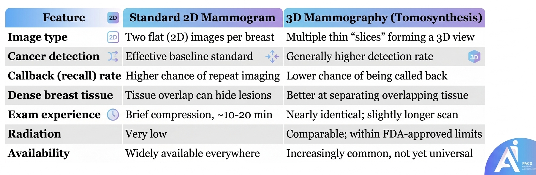
About this comparison: Availability and dose vary by center and protocol. The graphic’s dose comparison is not universal: DBT with separately acquired 2D images adds exposure, while synthetic 2D avoids that extra acquisition. A 3D examination still involves compression and can lead to a callback.
Who Benefits Most from 3D Mammography?
DBT reduces overlapping tissue and can improve cancer detection and reduce callbacks compared with 2D mammography in screening studies. The benefit varies by breast density, age and screening setting. Dense tissue can still hide a cancer. Read more about how dense breast tissue affects mammogram results. Your clinician can assess whether additional imaging is appropriate for your risk and findings.
Diagnosis & What to Expect During Your Exam
Preparing for either exam is straightforward. On the day of your appointment, avoid using deodorant, antiperspirant, powders, or lotions on your chest and underarms, because metallic particles in these products can appear as spots on the images. Wearing a two-piece outfit makes undressing from the waist up easier.
During the test, a technologist will position each breast on the imaging plate and apply compression for a few seconds. Compression can be uncomfortable or painful. Tell the technologist if it hurts so they can help with positioning. The actual imaging takes only moments.
A radiologist—a physician trained to interpret medical images—reviews your results and reports findings using a standardized scale called BI-RADS. If something needs a closer look, you may be asked to return for additional imaging such as a diagnostic mammogram or ultrasound. Being recalled does not mean you have cancer; most callbacks turn out to be benign.
Accuracy, Recall Rates, and Radiation
Screening studies have found higher cancer detection and fewer recalls with DBT than with 2D mammography alone. Results vary by population and protocol. Neither technique detects every cancer, and a callback does not itself mean cancer.
Radiation dose depends on the equipment and protocol. DBT with separately acquired 2D images adds exposure. A synthetic 2D image is reconstructed without another exposure, but the total dose is not always identical to a standard 2D examination. Ask your imaging center which approach it uses. ACR/RSNA patient information on tomosynthesis.
Is Tomosynthesis the Same as CT or MRI?
No. Breast tomosynthesis reconstructs slices from X-rays taken over a limited arc while the breast is compressed. It is not a conventional CT scan. MRI uses magnetic fields and radio waves and has different clinical uses. Screening and diagnostic describe why an exam is performed; 2D and 3D describe the imaging technique. A 3D mammogram can be used for screening or diagnostic assessment.
Practical Tips for Patients
- Ask what your facility offers. Not every clinic has 3D equipment. If you have dense breasts or a strong family history, ask whether tomosynthesis is available to you.
- Know your breast density. If your report or notification mentions dense tissue, discuss whether 3D mammography or additional imaging (such as ultrasound or MRI) is appropriate.
- Check coverage in advance. Ask your imaging center and insurer about availability, coverage and any out-of-pocket cost.
- Bring prior images. Comparing this year’s exam with previous ones helps the radiologist spot subtle changes and reduces unnecessary callbacks.
- Stay on schedule. Follow the screening plan agreed with your clinician and attend any recommended diagnostic follow-up.
- Don’t panic over a callback. Being asked to return for more imaging is common and usually ends with reassuring news.
Already have a report? Our Mammogram Second Opinion Online Service explains how an independent review works. It may agree with the original report or suggest clarification; a different diagnosis is not guaranteed. If no report has been issued, see online radiology reporting. Confirm that your complete original DICOM examination, including the full DBT series when applicable, can be reviewed before ordering. Screenshots or a report PDF alone are not a substitute for the image series.
Evidence and Screening Guidance
A systematic review published in European Radiology in 2022 found higher cancer detection and fewer recalls with DBT plus synthetic 2D images than with digital mammography alone. It found no difference in pooled interval-cancer rates, with limited data for that outcome. Better detection does not by itself establish a reduction in breast-cancer deaths. Read the systematic review.
The 2024 USPSTF recommendation recognizes both digital mammography and DBT as effective screening options. It recommends mammography every two years from ages 40 to 74 for its defined screening population. People with a previous breast cancer, certain high-risk genetic syndromes or other high-risk histories need a separate plan. Its insufficient-evidence statement about additional ultrasound or MRI after a negative mammogram in dense breasts should not be confused with a recommendation against DBT. Read the USPSTF recommendation.
Conclusion
Both 2D mammography and DBT are established screening options. The choice depends on your circumstances and local availability. If you have a new breast symptom or an abnormal result, arrange the recommended clinical assessment rather than waiting for your next routine screening visit. An online review should not delay recommended diagnostic imaging or biopsy.