

What Is an Abnormal Mammogram?
An abnormal mammogram identifies a finding that needs assessment, such as a mass, calcifications, distortion, or asymmetry. The word “abnormal” alone does not establish whether cancer is present. Read the BI-RADS assessment and recommendation in your report: an incomplete screening result and a suspicious diagnostic finding need different follow-up.
After screening, the radiologist may need additional views, an ultrasound, or previous images before making a final assessment. Overlapping tissue and dense breasts can make interpretation harder. A callback should be completed even when the finding may turn out to be benign.
If you already have a report and want another interpretation, an independent review can assess the available images and explain whether it agrees with the reported findings. It cannot replace an examination, missing diagnostic views, or a recommended biopsy.
Learn how our Mammogram Second Opinion service works, including the images needed for review.
How Common Is a Callback?
A callback after screening is common. The American Cancer Society reports that fewer than 1 in 10 women called back after a screening mammogram are found to have cancer. That figure describes screening callbacks as a group; it does not describe the risk of a BI-RADS 4 or 5 finding.
Callbacks are more common at a first mammogram, when previous images are unavailable for comparison. Your own next step depends on the actual finding and the completed diagnostic assessment.
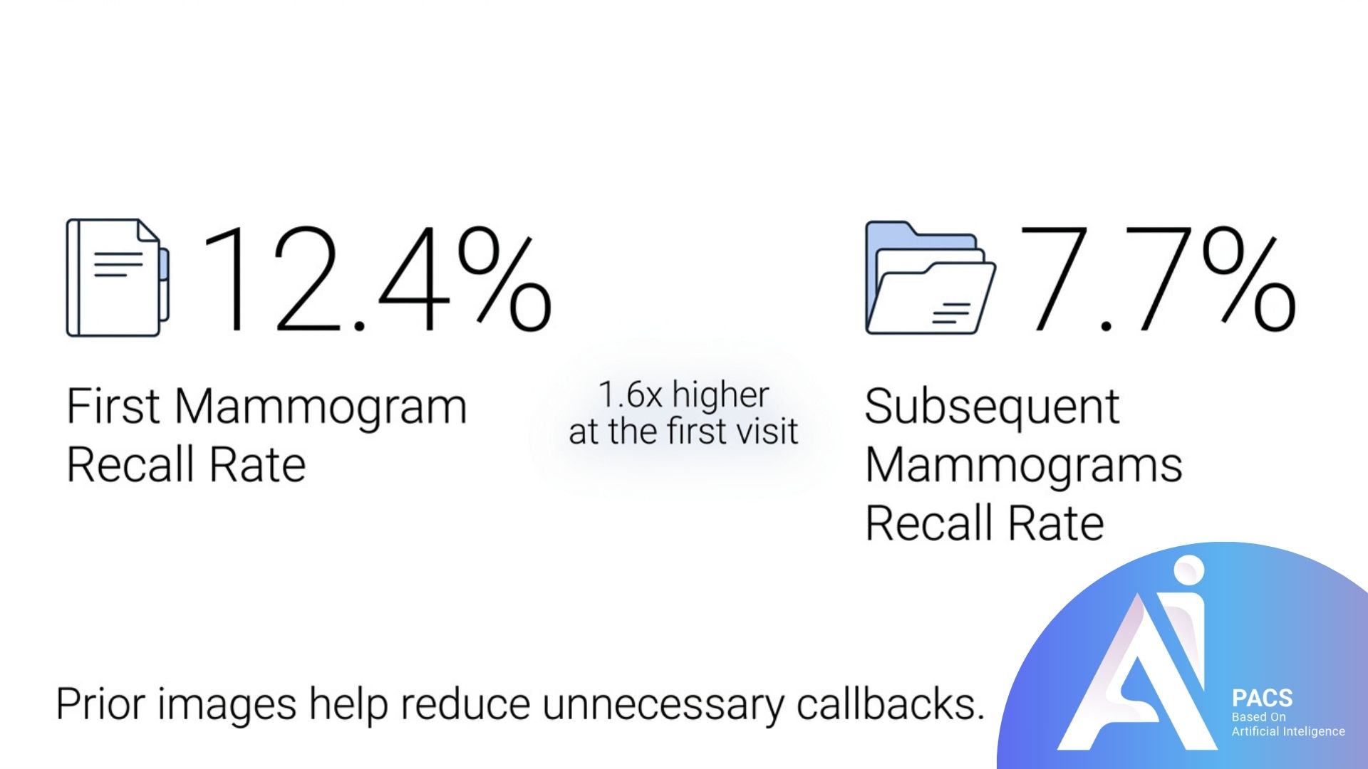
Ask your previous imaging center to share earlier mammograms. Comparing images over time can help the radiologist determine whether a finding is new or stable.
Understanding Your BI-RADS Score
Radiologists describe mammogram results using a standardized scale called BI-RADS (Breast Imaging Reporting and Data System). Knowing your number helps you understand exactly what your abnormal mammogram means and what to expect next.
BI-RADS 0 — Incomplete
BI-RADS 0 means the assessment is incomplete. Additional imaging and/or comparison with earlier mammograms is needed. It is not a final cancer-risk category.
BI-RADS 3 — Probably Benign
BI-RADS 3 means probably benign, with no more than a 2% likelihood of cancer. Short-interval imaging follow-up is usually recommended, often at six months. Follow the interval in your report rather than arranging an immediate repeat solely because the word “abnormal” appears.
BI-RADS 4 and 5 — Biopsy Usually Recommended
BI-RADS 4 is suspicious and spans a range of concern, described by subcategories 4A, 4B, and 4C. BI-RADS 5 is highly suggestive of malignancy, with a likelihood of at least 95%. A biopsy is generally recommended for both categories. Do not apply reassuring screening-callback statistics to these findings or delay the recommended assessment while seeking another opinion.
Read for more information: What Does BIRADS Mean in Mammography?
What Tests Come Next?
Once you are recalled, your care team will tailor the next steps to what they saw. Here is what typically happens.
Diagnostic Mammogram
This is a more detailed mammogram focused on the area of concern. A follow-up diagnostic mammogram with spot compression views is often ordered, frequently alongside a diagnostic ultrasound, to clarify the finding. The result is then resolved into a clear category.
Breast Ultrasound
Ultrasound uses sound waves and no ionizing radiation. It can help assess whether a finding contains fluid or is solid and may guide a biopsy. Ultrasound findings are interpreted together with the mammogram and clinical information.
Breast MRI
In selected cases—such as high-risk patients or unclear findings—an MRI provides highly detailed images. The same BI-RADS categories apply, though the recommended next steps may differ slightly.
Biopsy
A biopsy takes a tissue sample for laboratory examination when a finding needs tissue diagnosis. Your care team chooses the sampling method and imaging guidance appropriate to the area. An online image review does not replace pathology or a biopsy recommended by your treating team.
How to Prepare and What to Expect
Preparing for your follow-up appointment can make the experience smoother and less stressful. A little planning goes a long way.
Bring previous mammograms and reports, or arrange for your imaging center to transfer them. Tell the team about breast symptoms and prior procedures. Follow the facility’s preparation instructions; avoid deodorant, powder, or lotion on your breasts and underarms on the day of mammography.
It is also wise to sort out logistics early. Contacting your insurance provider before a diagnostic appointment helps you understand expected out-of-pocket costs, and many imaging centers have financial counselors who can help navigate questions or assistance options. Finally, consider bringing a trusted friend or family member for support.

Why Acting Promptly Matters
Contact the imaging center or referring clinician to clarify your report’s recommended next step and timing. BI-RADS 0 requires completion of the assessment; BI-RADS 3 usually calls for a planned short-interval check; suspicious findings require discussion of biopsy.
If you have not received your report, ask when it will be available and how you will receive it. Do not assume that no news means a normal result. Report a new or persistent lump or other breast change to your clinician, even if imaging was described as normal.
Keep appointments already recommended by your care team while arranging any independent image review. If access or scheduling is difficult, tell the referring clinician so they can help coordinate the next step.
Research Context
Research describes outcomes across groups, not the diagnosis or safe waiting time for one person. Two studies help explain why completing follow-up and interpreting callback statistics carefully matter.
Oluyemi and colleagues reported follow-up patterns after nearly 2.9 million abnormal screening mammograms in the Journal of the American College of Radiology in 2024. Among documented follow-up, the median interval to diagnostic evaluation was nine days. This was an observational finding, not a recommended deadline or a promise about service turnaround.
Ho and colleagues’ 2022 JAMA Network Open study compared cumulative false-positive outcomes over ten years of screening with tomosynthesis and digital mammography. Its outcomes included false-positive recalls and biopsy recommendations—not simply completed benign biopsies. Screening history, age, density, and screening interval affect these estimates; they cannot determine whether an individual abnormality is benign.
Calcifications are assessed by their appearance and distribution. A general benign-outcome percentage cannot replace the BI-RADS assessment of a particular pattern.
Tomosynthesis (3D mammography) can reduce callbacks and improve detection in screening populations, according to RadiologyInfo.org. It does not eliminate the need to investigate a finding already identified.
Conclusion
An abnormal mammogram needs a clear follow-up plan. Check the BI-RADS category, the recommended test, and the timeframe with your care team. A callback is not itself a cancer diagnosis, but a suspicious finding must not be dismissed using general screening statistics.
For an independent review, have the original DICOM images, the existing report, and prior imaging available if possible. A screenshot or a report PDF alone is not a substitute for diagnostic image review. Continue the care already recommended by your clinician.
Scientific References
- Oluyemi ET, Grimm LJ, Goldman L, et al. Rate and Timeliness of Diagnostic Evaluation and Biopsy After Recall From Screening Mammography in the National Mammography Database. J Am Coll Radiol. 2024;21(3):427-438. doi:10.1016/j.jacr.2023.09.002.
- Ho TH, Bissell MCS, Kerlikowske K, et al. Cumulative Probability of False-Positive Results After 10 Years of Screening With Digital Breast Tomosynthesis vs Digital Mammography. JAMA Network Open. 2022;5(3):e222440. doi:10.1001/
jamanetworkopen. 2022.2440. - American Cancer Society. Understanding Your Mammogram Report.
- Johns Hopkins Medicine. Understanding Your Mammogram Report.
- Cleveland Clinic. BI-RADS (Breast Imaging-Reporting & Data System). Last updated 10/23/2024.
- D’Orsi CJ, Sickles EA, Mendelson EB, Morris EA, et al. (Eds). ACR BI-RADS Atlas, Breast Imaging Reporting and Data System. American College of Radiology, 2013.
- RadiologyInfo.org. Mammography.
- National Cancer Institute (NIH). Mammograms Fact Sheet.
- U.S. FDA. Mammography: What You Need to Know.
- American Cancer Society. Getting Called Back After a Mammogram.