

Asymmetry in Mammogram: Understanding Its Implications and Importance
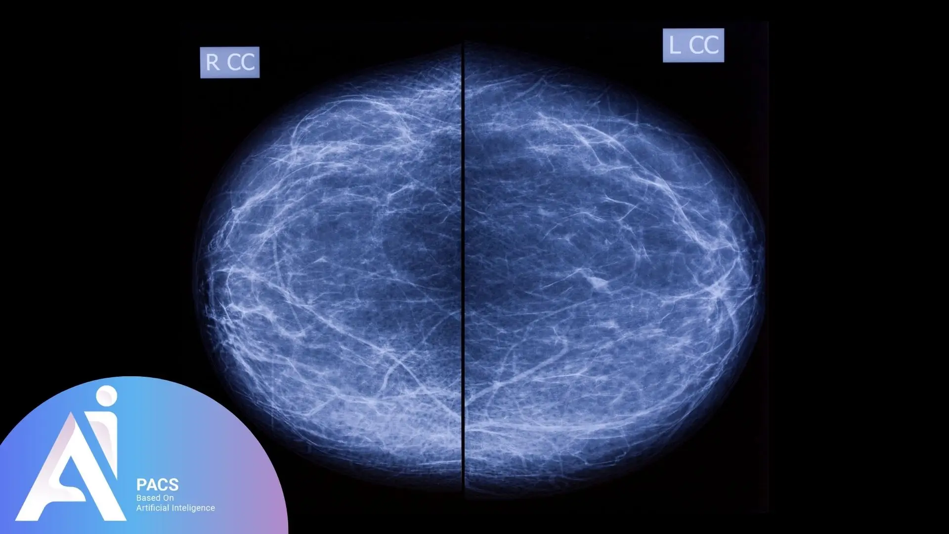
Asymmetry in mammogram findings indicates a difference in density, structure, or size between corresponding areas of the two breasts. While it can signal benign conditions like anatomical variation or hormonal changes, it may also require further investigation for potential issues such as cysts, fibroadenomas, or malignancies. Radiologists use additional imaging techniques to assess the significance of this asymmetry and decide whether additional imaging or follow-up is needed.
Asymmetry terms you may see in a mammogram report
- Asymmetry (seen on one view)
An area of increased density is seen on one mammographic view. Additional views may help determine whether it reflects overlapping tissue or a finding that needs further assessment. The word asymmetry alone does not establish that a finding is harmless. - Focal Asymmetry
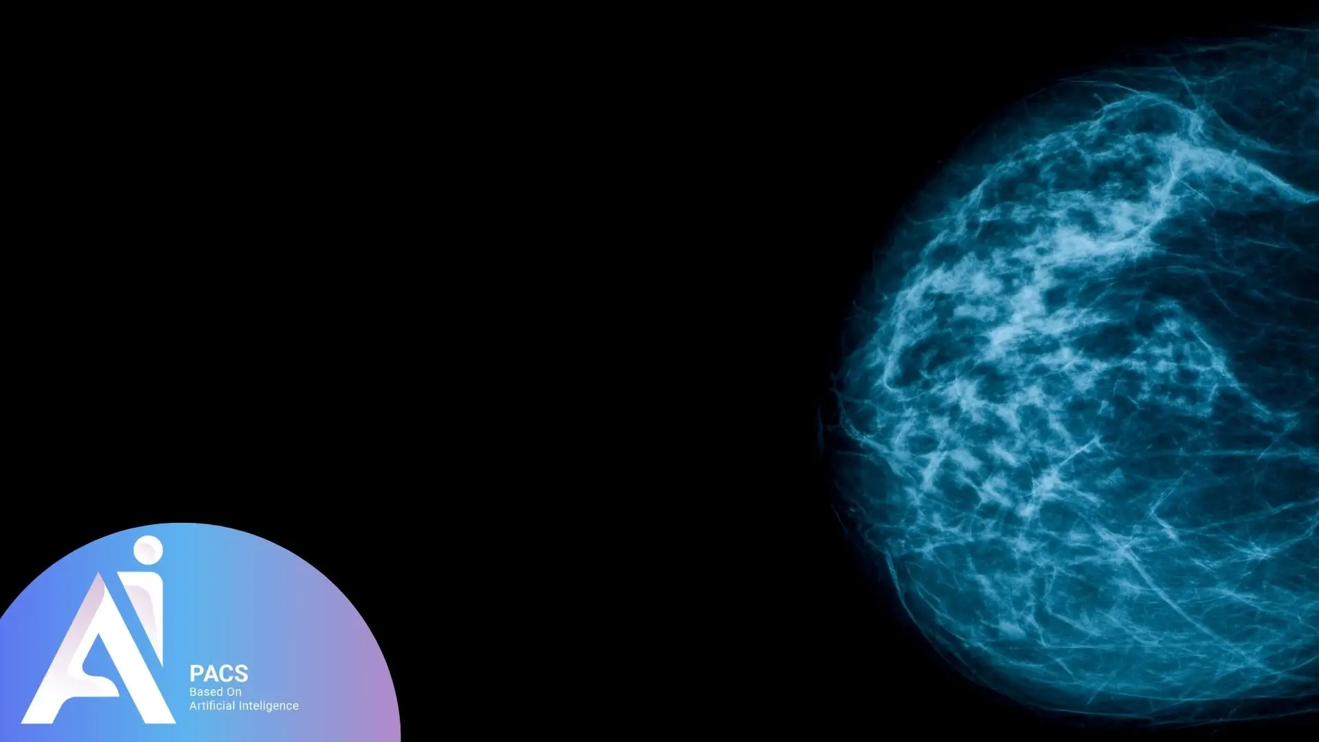
Focal asymmetry appears as a localized area of denser breast tissue visible in two different mammographic views. To rule out concerning causes, additional imaging, such as spot compression mammography or ultrasound, may be required. - Global Asymmetry
Global asymmetry is a more extensive density difference in a larger area of one breast than the other. This type is often associated with normal breast tissue variation but might need further evaluation if it is a new finding. - Developing Asymmetry
This describes an area that is new, larger, or more conspicuous compared with earlier mammograms. Reports may use the term developing asymmetry to describe this change. The radiologist considers the complete findings and may recommend additional imaging or biopsy.
Each type of asymmetry requires a tailored approach to evaluation and follow-up to determine whether it represents a benign variation or a potential health concern.
Common Causes of Asymmetry in Mammograms
Non-cancerous causes
- Breast Density Variations
Differences in breast tissue density are common and often attributed to genetic factors or hormonal changes. Dense tissue can create asymmetry, especially in younger women. - Hormonal Changes
Hormonal fluctuations during the menstrual cycle, pregnancy, or menopause can temporarily alter breast tissue, leading to asymmetry in imaging. - Fibrocystic Changes
These benign changes result in lumpiness or uneven texture in the breast tissue, which may appear asymmetric on a mammogram. - Post-Surgical or Radiation Effects
Scars or tissue changes from previous breast surgeries, biopsies, or radiation therapy can cause localized asymmetry. - Normal Anatomical Variations
Slight differences in size, shape, or tissue distribution between breasts are natural and do not usually indicate health concerns.
Potential pathological causes
- Benign Tumors
Growths like fibroadenomas or lipomas may cause localized asymmetry, though they are typically non-cancerous and easily managed. - Breast Cysts
Fluid-filled sacs within the breast can create density differences, especially if they are large or concentrated in one area. - Inflammation or Infection
Conditions such as mastitis or abscesses may lead to swelling or tissue changes that appear asymmetric on a mammogram. - Developing Breast Cancer
Asymmetry, particularly a new or developing one, can be an early indicator of breast cancer, including ductal carcinoma in situ (DCIS) or invasive breast cancer. - Other Malignant Lesions
Rare conditions, such as phyllodes tumors or metastatic lesions, might also manifest as asymmetry and require immediate attention.
How Radiologists Evaluate Asymmetry in Mammogram Findings
Diagnostic tools and techniques
Radiologists use advanced imaging tools to assess asymmetry in mammogram findings. If asymmetry is detected, additional imaging techniques like spot compression or magnification views are employed for clearer images, helping to rule out artifacts or overlapping tissue.
Breast ultrasounds are typically used next to differentiate between solid masses, cysts, or normal tissue. For complex cases, breast MRI provides detailed three-dimensional imaging that can uncover abnormalities not seen in traditional mammograms.
If suspicious findings arise, a biopsy is recommended to obtain a tissue sample for analysis. Comparing current mammograms with previous ones is essential to determine whether the asymmetry is new, stable, or worsening, guiding further evaluation.

BI-RADS categorization and its role in assessment
BI-RADS 0 means the assessment is incomplete: additional imaging and/or comparison with previous studies is needed. For explanations of all categories, read our guide to BI-RADS 0–6.
The Breast Imaging-Reporting and Data System (BI-RADS) is a standardized classification used by radiologists to evaluate breast findings, including asymmetry:
– BI-RADS 1 and 2 indicate negative or benign findings; follow the screening plan recommended for your age, symptoms, and risk.
– BI-RADS 3 suggests probably benign asymmetry, recommending short-term follow-up imaging.
– BI-RADS 4 is assigned to suspicious findings, typically warranting a biopsy, and further categorized into subcategories (4A, 4B, 4C) based on the level of suspicion.
– BI-RADS 5 indicates findings highly suggestive of malignancy, requiring immediate action.
– BI-RADS 6 is used when cancer has already been confirmed by biopsy; it is not a diagnosis made from the mammogram alone.
This system promotes clear communication among radiologists and healthcare providers for accurate diagnoses and effective patient care.
Questions to ask about your own mammogram
- Where is the asymmetry, and was it seen on one view or more?
- Was it present on earlier mammograms, or has it changed?
- What BI-RADS assessment and follow-up were recommended?
If you want an independent assessment of the images behind your report, our mammogram second-opinion service explains the review process. Provide the complete original DICOM images, your report, and previous studies when available. A report excerpt or screenshot alone cannot replace an image review. A second opinion may agree with the original report and should not delay recommended diagnostic imaging or biopsy.
Does Asymmetry in Mammogram Findings Always Indicate Cancer?
Asymmetry in mammogram findings does not always indicate cancer. While it can be a sign of malignancy in some cases, numerous benign causes are far more common. The key to understanding asymmetry lies in differentiating between benign and malignant causes through careful evaluation.
Benign causes
Asymmetry in mammograms is often due to non-cancerous factors. Normal variations in breast density or tissue distribution can cause asymmetric findings, while hormonal changes during the menstrual cycle, pregnancy, or menopause may lead to temporary breast tissue alterations. Additionally, benign conditions like cysts, fibroadenomas, or scar tissue can create areas that mimic asymmetry. Radiologists assess these possibilities using the imaging features and clinical context. Additional imaging or a biopsy may be needed when the finding is uncertain.
Malignant causes
Asymmetry in breast tissue can indicate an underlying malignancy, especially if it is new or different from previous mammograms. When linked to conditions like ductal carcinoma in situ (DCIS) or invasive breast cancer, asymmetry may also show other warning signs, such as architectural distortion, microcalcifications, or a palpable lump. Developing asymmetry is particularly concerning and requires prompt follow-up with additional imaging or a biopsy to confirm or rule out cancer.
When to Be Concerned About Asymmetry in Mammogram Findings
Asymmetry in mammogram findings is not always a cause for alarm, but certain situations warrant closer attention and further evaluation. Understanding when to be concerned can help patients seek timely medical advice and ensure early detection of potential issues.
Developing Asymmetry
A newly developed asymmetry compared to previous mammograms is often considered a red flag. This type of asymmetry could indicate changes in breast tissue caused by underlying conditions, including malignancies. Radiologists typically recommend additional imaging or a biopsy to determine the cause.
Associated Suspicious Features
Concern increases when other suspicious findings accompany asymmetry. These may include architectural distortion, the presence of microcalcifications, or a palpable lump in the breast. Changes in skin texture, nipple retraction, or unusual discharge are warning signs that necessitate immediate follow-up.
Persistent Focal Asymmetry
The focal asymmetry that persists across multiple views and imaging sessions may require additional diagnostic tests, such as spot compression, ultrasound, or breast MRI. Although it may be benign, the persistence of asymmetry without a clear explanation often calls for further investigation.
Symptoms or Risk Factors
If asymmetry is identified alongside symptoms like breast pain, swelling, or visible changes, or if the patient has a family history of breast cancer or genetic predisposition (e.g., BRCA mutations), a more thorough evaluation is essential.
Early follow-up with a healthcare provider is critical to determine the cause of the asymmetry and ensure appropriate treatment. Regular screenings and timely interventions are vital in effectively addressing potential concerns.
Screening and Follow-Up After an Asymmetry Finding
Routine screening and diagnostic follow-up are different. If your report recommends additional views, ultrasound, or biopsy, ask your care team when to arrange it rather than waiting for your next routine screening appointment.
Routine screening guidance
Screening schedules depend on your country, age, and individual risk. For example, the US Preventive Services Task Force recommends mammography every two years for women ages 40–74 within the population covered by its recommendation. People with certain inherited cancer-risk syndromes or a personal history of breast cancer need an individualized plan. Discuss the appropriate schedule with your clinician.
Can lifestyle changes make the finding disappear?
A healthy lifestyle supports general health, but it cannot determine whether an asymmetry is benign or replace recommended diagnostic follow-up. Share new breast symptoms and relevant medical history with your clinician, and obtain earlier mammograms for comparison when available.
Conclusion
Asymmetry describes an imaging finding, not a cancer diagnosis. Its significance depends on the images, prior examinations, and the final BI-RADS assessment. Ask what was seen and which follow-up is recommended. If you request an independent image review, provide the original DICOM studies and report, and keep your planned clinical appointments.
References:
American Cancer Society: Understanding Your Mammogram Report