
Magnetic Resonance Imaging (MRI) is a powerful diagnostic tool that uses magnetic fields and radio waves to create detailed images of the body’s organs and tissues. When contrast dye is added to an MRI, it significantly enhances the clarity and detail of these images, particularly for the brain. This advanced technique allows doctors to visualize structures and identify subtle abnormalities that might otherwise go unnoticed.
A Brain MRI with contrast is a specialized imaging test that provides an incredibly detailed view of your brain’s anatomy and blood supply. It’s often recommended when a standard MRI isn’t sufficient to pinpoint a diagnosis or to get a clearer picture of a known condition.
What is a Contrast Agent and How Does It Work?
A contrast agent, often referred to as “dye,” is a substance injected into your bloodstream during the MRI scan. For brain MRIs, the most common contrast agent is gadolinium-based.
Here’s how it works:
- Magnetic Properties: Gadolinium is a paramagnetic substance. This means it alters the magnetic properties of nearby water molecules in your body when exposed to the strong magnetic field of the MRI scanner.
- Signal Enhancement: This alteration causes the MRI scanner to pick up a stronger signal from areas where the contrast agent accumulates.
- Highlighting Abnormalities: Diseased or abnormal tissues, such as tumors, areas of inflammation, or active infections, often have a different blood supply or a “leaky” blood-brain barrier compared to healthy tissue. The contrast agent accumulates in these abnormal areas, making them appear brighter (enhanced) on the MRI images. This enhancement helps radiologists distinguish between normal and abnormal tissue, delineate the precise location and extent of a lesion, and assess its characteristics.
- Vascular Visualization: Contrast also significantly improves the visualization of blood vessels, helping to identify blockages, aneurysms, or other vascular malformations.
Why is a Contrast Brain MRI Performed?
A contrast-enhanced Brain MRI is a crucial diagnostic tool used for a variety of neurological conditions. It is particularly valuable when:
1. Detecting and Characterizing Brain Tumors
- Identification: Tumors, whether primary (originating in the brain) or metastatic (spread from elsewhere), often have abnormal blood vessels or are located in areas where the blood-brain barrier is disrupted. The contrast agent highlights these areas, making tumors more visible.
- Differentiation: It helps radiologists differentiate between different types of tumors (e.g., benign vs. malignant) and determine their exact size, shape, and extent.
- Monitoring Treatment: Contrast MRIs are used to monitor how well a tumor is responding to treatment, such as chemotherapy or radiation therapy, by showing changes in tumor size or enhancement patterns.
2. Evaluating Inflammation and Infections
- Inflammatory Conditions: Diseases like Multiple Sclerosis (MS) cause inflammation in the brain. Active MS lesions often enhance with contrast, indicating areas of current inflammation.
- Infections: Brain abscesses, meningitis (inflammation of the membranes surrounding the brain and spinal cord), or encephalitis (inflammation of the brain itself) can be better visualized and localized with contrast. The contrast agent can highlight the inflamed tissues or collections of pus.
3. Assessing Blood Vessel Abnormalities
- Aneurysms and Arteriovenous Malformations (AVMs): While dedicated MRA (Magnetic Resonance Angiography) sequences are often used, contrast can supplement the evaluation of these vascular structures by showing blood flow and the extent of abnormalities.
- Vasculitis: Inflammation of the blood vessels in the brain can be detected and assessed using contrast-enhanced MRI.
4. Investigating Seizures and Headaches
- Underlying Causes: For persistent or unexplained seizures and severe headaches, a contrast MRI can help identify underlying structural causes like small tumors, vascular malformations, or areas of scarring that might trigger these symptoms.
5. Evaluating Changes After Stroke or Injury
- Ischemic Stroke: In some cases, contrast can help identify areas of acute inflammation or breakdown of the blood-brain barrier in the early stages of an ischemic stroke, although other MRI sequences (like diffusion-weighted imaging) are primary for detecting acute stroke.
- Traumatic Brain Injury (TBI): Contrast can help identify areas of bleeding or inflammation resulting from head trauma that might not be apparent on a non-contrast scan.
6. Pre-Surgical Planning
- Precise Localization: For neurosurgeons, a contrast MRI provides critical information about the exact location, size, and relationship of a lesion to surrounding healthy brain tissue and important blood vessels, aiding in surgical planning.

What to Expect During Your Scan
Undergoing a Brain MRI with contrast is a straightforward process, designed to be as comfortable as possible.
Before the Scan
- Consultation: Your doctor will discuss why the MRI is needed and review your medical history. It’s crucial to inform your doctor if you:
- Have any allergies, especially to contrast agents or medications.
- Have kidney problems (as contrast is filtered by the kidneys).
- Are pregnant or breastfeeding.
- Have any metal implants or foreign bodies (though most are MRI-safe, it’s essential to confirm).
- Are taking certain medications.
- Preparation: You may be asked to avoid eating or drinking for a few hours before the scan, especially if sedation is planned (though rare for routine brain MRIs). You’ll change into a hospital gown.
- Screening: You’ll be asked a series of questions to ensure there are no contraindications for the MRI, such as certain types of pacemakers or metal fragments in your eyes.
During the Scan
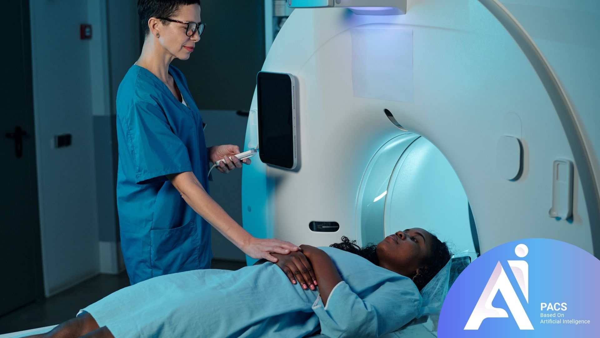
- Positioning: You will lie down on a movable table that slides into the opening of the MRI scanner, which looks like a large tube. You’ll be positioned comfortably, usually on your back, and a special head coil (a device that helps capture clear images) might be placed around your head.
- Contrast Injection: An intravenous (IV) line will be inserted into a vein in your arm or hand. The contrast agent will be injected through this line either manually by a technologist or automatically by a power injector at specific times during the scan. You might feel a cool sensation or a metallic taste in your mouth during the injection.
- Scanning: The MRI machine produces loud tapping or banging noises during operation. You’ll be given earplugs or headphones to reduce the noise. It’s essential to remain as still as possible throughout the scan to ensure the images are clear and free from motion artifact. The technologist will communicate with you through an intercom system.
- Duration: A Brain MRI with contrast typically takes about 30 to 60 minutes to complete, depending on the specific sequences required.
After the Scan
- Recovery: You can usually resume your normal activities immediately after the scan.
- Hydration: It’s often recommended to drink extra fluids for the rest of the day to help your body flush out the contrast agent.
- Side Effects: While rare, some people might experience mild side effects like nausea, headache, or dizziness. Allergic reactions are very uncommon but can range from mild (rash) to severe (anaphylaxis). Radiologists and technologists are trained to manage these reactions.
Understanding the Results
Once the scan is complete, a radiologist—a physician specifically trained in interpreting medical images—will carefully review all the images. If you’d like an independent read, you can have your MRI read online by an expert radiologist.
Normal Findings
In a healthy brain, the MRI with contrast will show:
- Clear brain structures with no abnormal areas of enhancement.
- Normal-appearing blood vessels without any bulges or blockages.
- No signs of inflammation, infection, or masses.
Abnormal Findings
When abnormalities are present, the contrast agent will highlight them:
- Tumors: Appear as bright, enhancing masses, often with irregular shapes, indicating abnormal blood supply. The radiologist can assess their location, size, and whether they are pushing on or invading surrounding brain tissue.
- Inflammation (e.g., MS plaques): May show as distinct, often ring-like, areas of enhancement in the white matter of the brain.
- Infections (e.g., Abscess): Typically appear as a collection of pus that enhances brightly around its edges, with a possible area of decreased signal in the center.
- Vascular Issues: Areas of abnormal blood flow or vessel wall enhancement might indicate conditions like vasculitis or certain types of stroke-related changes.
- Scar Tissue: Old injuries or surgeries might leave scar tissue that can enhance with contrast.
The radiologist will compile a detailed report of their findings, which will be sent to your referring physician. Your doctor will then discuss the results with you and explain what they mean in the context of your overall health and symptoms.
If findings are complex, using AI PACs MRI Second Opinion Service can provide additional reassurance and expert review.
Latest Scientific Insights: Advancements in Contrast MRI
Research continues to refine the use and interpretation of contrast-enhanced MRI for neurological conditions. Recent studies focus on:
- Quantitative Enhancement: Developing methods to precisely measure the degree of contrast enhancement, which can provide more objective information about tumor aggressiveness or treatment response. (Source: Journal of Magnetic Resonance Imaging, 2023)
- Gadolinium Retention: Ongoing research investigates the long-term effects of gadolinium-based contrast agents, including potential retention in the brain. While current evidence suggests it poses minimal risk for most patients, research is exploring ways to minimize or monitor this. (Source: Radiology, 2022)
Conclusion
A Brain MRI with Contrast is an invaluable tool that provides unparalleled detail of the brain’s structure and function. By highlighting abnormalities with a contrast agent, it empowers physicians to diagnose a wide range of conditions—from tumors and infections to inflammatory diseases and vascular issues—with greater accuracy. While the procedure involves lying in a scanner and receiving an injection, it is generally safe and offers crucial insights that guide effective treatment strategies, ultimately leading to better patient outcomes.
Scientific References Used
- RadiologyInfo.org: (Accessed May 2, 2026) – Comprehensive patient information on MRI of the brain.
- Mayo Clinic: (Accessed May 2, 2026) – General overview of MRI.
- Cleveland Clinic: (Accessed May 2, 2026) – Information on MRI procedures and uses.
- National Institute of Biomedical Imaging and Bioengineering (NIBIB): (Accessed May 2, 2026) – Scientific explanation of MRI technology.
- PubMed (Example search for recent insights): Searching for “contrast-enhanced brain MRI tumors review” or “gadolinium retention MRI” yields numerous studies. (Accessed May 2, 2026)
- Radiopaedia: MRI contrast agents (Accessed May 2, 2026) – Detailed radiological perspective on contrast agents.