
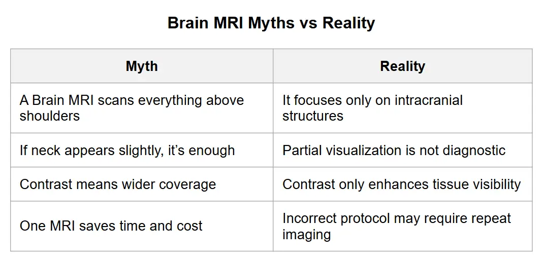
Magnetic Resonance Imaging (MRI) is one of the most advanced imaging tools in modern medicine. Many patients wonder whether a Brain MRI also captures the neck region. The short answer is: No, a routine Brain MRI does not cover the neck. However, depending on your symptoms or physician orders, additional imaging may be needed.
What Is a Brain MRI?
A Brain MRI is a non‑invasive test that uses magnetic fields and radio waves to produce detailed images of the brain, cerebellum, brainstem, and sometimes the uppermost cervical spinal cord. It does not use radiation. According to RadiologyInfo.org (2024), MRI is essential for diagnosing tumors, stroke, inflammation, and neurological disorders.
What Area Does a Standard Brain MRI Cover?
A standard Brain MRI covers the top of the skull down to the base of the skull. In most cases, it includes only a small part of the upper cervical spine (C1–C2), which is not enough for diagnosing neck problems.
Why Part of the Neck Sometimes Appears
Some upper neck structures appear unintentionally because the brainstem transitions into the spinal cord. However, this partial view is not diagnostic.
What Is a Cervical Spine MRI?
If neck-related symptoms exist, doctors typically order a Cervical Spine MRI. This scan evaluates cervical vertebrae (C1–C7), discs, spinal cord, nerve roots, and soft tissues. According to Mayo Clinic (2023), it is the best imaging method for disc herniation, spinal cord compression, and degenerative neck disease.
When the neck itself is the focus, a dedicated neck study is used instead — our guide on how to read a neck CT scan explains what those images show.
When Do Doctors Order Both Brain and Neck MRI?

Doctors may order both scans together in cases such as:
- Multiple Sclerosis (MS)
- Trauma involving head and neck
- Tumor staging
- Neurological symptoms like arm numbness or weakness
The 2023 McDonald Criteria (Lancet Neurology) emphasize combined brain and spine MRI for early MS diagnosis.
If your scan was ordered for multiple sclerosis, our guide to what an MS brain MRI report says explains the terms you may see.
Why a Brain MRI Cannot Replace a Neck MRI
Brain MRI uses a head coil and sequences optimized for brain tissue. Cervical Spine MRI uses a neck coil and sequences optimized for vertebrae, discs, and nerve roots. According to Radiopaedia (2024), proper imaging requires targeted protocols, so one scan cannot replace the other.
Symptoms Suggesting You Need a Neck MRI
You may need a Cervical Spine MRI if you experience:
- Neck pain
- Pain radiating to arms
- Hand weakness or tingling
- Shoulder and arm numbness
- Electric shock sensation when bending the neck
On the other hand, Brain MRI is more suitable for headaches, seizures, memory problems, and stroke symptoms. If you already have a cervical spine MRI, you can request a neck MRI second opinion from a subspecialty radiologist.
Practical Recommendations Before Scheduling Your MRI
- Clearly describe your symptoms to your doctor.
- Ask whether you need Brain MRI, Cervical MRI, or both.
- Bring previous imaging to avoid unnecessary scans.
- Consider getting a radiology second opinion if unsure. If you already have the images, you can send them for online MRI reporting by a board-certified radiologist.
In complex cases, especially when symptoms overlap between brain and cervical spine conditions, obtaining an expert radiology review may help avoid unnecessary repeat scans. If you have already undergone a Brain MRI, you may also consider requesting a professional consultation or a second opinion on your MRI results to ensure the findings have been accurately interpreted and that the correct imaging protocol was used. This can help clarify whether additional imaging—such as a Cervical Spine MRI—is truly necessary.
Latest Scientific Findings (2023–2025)
- The 2023 McDonald Criteria highlight the importance of combined brain and spine MRI for early MS diagnosis.
- A 2024 Radiology (RSNA) study shows that AI tools improve MRI protocol selection based on symptoms.
- A 2023 AJNR study found that dedicated cervical MRI detects spinal cord compression far better than partial field images from a Brain MRI.
Common Patient Misconceptions
Conclusion
A routine Brain MRI does not include the neck. It focuses on the brain and uppermost spinal cord. For diagnosing neck pain, disc issues, or nerve compression, a dedicated Cervical Spine MRI is required. Selecting the right imaging test ensures faster diagnosis and helps avoid unnecessary repeat scans. If you are uncertain which imaging study is appropriate, you can request a radiology second opinion through AI-PACS radiology second opinion to receive expert guidance and ensure the correct scan is performed.
Scientific Sources Used
- RadiologyInfo.org (RSNA & ACR). Brain MRI. Updated 2024.
- Mayo Clinic. MRI Overview and Uses. 2023.
- Cleveland Clinic. Cervical Spine MRI. 2024.
- Radiopaedia.org. MRI Protocols – Brain and Spine. 2024.
- Thompson AJ et al. Diagnosis of Multiple Sclerosis: 2023 Revision of the McDonald Criteria. Lancet Neurology. 2023.
- American Journal of Neuroradiology. Cervical Myelopathy Imaging Accuracy. 2023.
- Radiology (RSNA). AI Optimization of MRI Protocols. 2024.