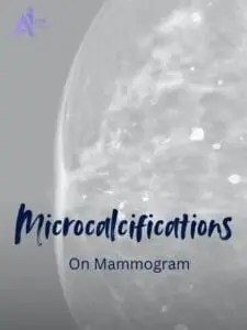

What are microcalcifications?
Microcalcifications are tiny deposits of calcium that form within breast tissue. These specks are often too small to feel during a physical exam but can be detected through imaging. While most microcalcifications are benign, specific patterns may indicate underlying conditions, including early signs of breast cancer.
Why are they detected during mammograms?
Mammograms are specialized imaging tools designed to detect subtle changes in breast tissue, including microcalcifications. The high-resolution images reveal these calcium deposits, allowing radiologists to evaluate their patterns and identify potential abnormalities that may require further investigation.
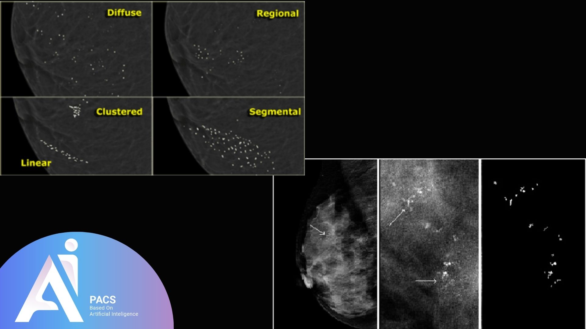
Types of Microcalcifications
Microcalcifications can be classified based on appearance, pattern, and clinical significance. Understanding these categories helps determine whether they are harmless or require further investigation.
Benign microcalcifications
Benign microcalcifications are typically associated with non-cancerous changes in breast tissue. These may result from aging, past injuries, fibrocystic changes, or calcification of milk ducts. They often appear as round, scattered, or evenly distributed specks on a mammogram and usually pose no threat.
Microcalcifications that need further assessment
Suspicious microcalcifications may suggest abnormal changes, such as ductal carcinoma in situ (DCIS) or invasive breast cancer. A cluster alone does not establish cancer. Shape, distribution, and changes from previous mammograms help determine whether additional views or a biopsy are needed.
For an independent image review, learn about our mammogram second-opinion service or start a mammogram review request. Provide the complete DICOM images, your report, and previous studies when available. A report or screenshot alone is not enough for diagnostic image interpretation.
Critical differences in patterns and appearance
The appearance and arrangement of microcalcifications play a crucial role in determining their significance:
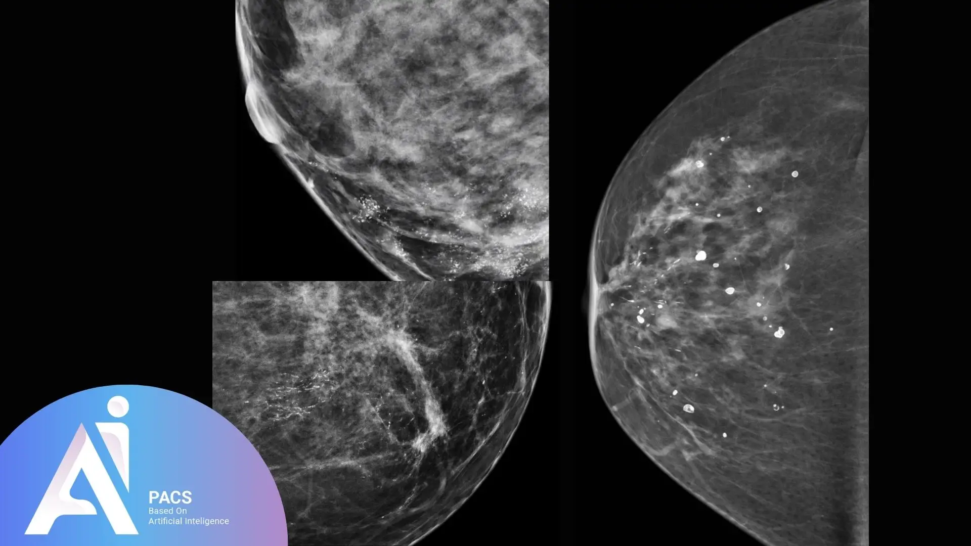
- Benign: Scattered, round, or smooth-edged deposits.
- Suspicious: Some irregular or fine linear patterns need further evaluation. Grouping alone is not a diagnosis. Radiologists assess the complete pattern and compare previous studies.
Causes of Microcalcifications
Microcalcifications in the breast can arise from various causes, most of which are benign. Common causes include natural aging processes, where calcium deposits form in milk ducts or breast tissue. Other benign conditions, such as fibrocystic changes, past injuries, or inflammation, can also lead to their development. In some cases, microcalcifications may signal ductal carcinoma in situ (DCIS), a non-invasive form of breast cancer, or other early malignancies. Factors like previous radiation therapy, calcified cysts, or residual calcifications from healing processes can also contribute. While most microcalcifications are harmless, their appearance and patterns are closely analyzed to rule out underlying issues.
How Microcalcifications Are Evaluated
Microcalcifications are evaluated using advanced imaging techniques to determine their nature and significance. Radiologists focus on their size, shape, distribution, and pattern to assess whether they are benign or suspicious, guiding further diagnostic steps if necessary.
Mammogram techniques for detecting microcalcifications
Mammograms are the primary tool for detecting microcalcifications. Digital mammography can show small calcium deposits in breast tissue. Specialized techniques like magnification views are often used to provide a closer, more detailed look at the microcalcifications, enabling a better assessment of their characteristics.
BI-RADS classification and its role
The BI-RADS (Breast Imaging Reporting and Data System) classification is a standardized tool radiologists use to describe mammogram findings, including microcalcifications. BI-RADS 0 means the assessment is incomplete; 1 is negative; 2 describes a benign finding; 3 usually requires short-interval follow-up. Categories 4 and 5 generally lead to a biopsy recommendation, while 6 means cancer has already been confirmed by biopsy. Follow the recommendation in your own report.
Read our guide to BI-RADS categories for the meaning of the assessment in your report.
Factors considered by radiologists
Radiologists consider several factors when evaluating microcalcifications:
- Pattern and distribution: The arrangement is assessed together with the shape of the deposits; a cluster can be benign or suspicious.
- Size and shape: Irregular shapes or varying sizes can raise concerns.
- Comparison: Previous mammograms help establish whether a finding is new or has changed. The full imaging assessment and medical history guide next steps.
When Are Microcalcifications a Concern?
The final assessment matters more than the word microcalcifications alone. Ask whether the finding is benign, probably benign, or suspicious, and whether more views are needed. A biopsy recommendation means tissue sampling is needed to clarify the finding; it does not itself confirm cancer.
Treatment and Follow-Up
Proper management of microcalcifications depends on their nature. They can range from benign findings requiring no treatment to precancerous or cancerous conditions needing intervention. Regular follow-up and monitoring play a crucial role in ensuring breast health.
Managing benign microcalcifications
Benign microcalcifications typically do not require treatment. These calcium deposits usually result from aging, previous injuries, or harmless tissue changes. Radiologists may recommend regular mammograms to monitor these findings over time and ensure no changes in their pattern or appearance.
If a biopsy is recommended
A mammography-guided needle biopsy can sample calcifications, including those not visible on ultrasound. A pathologist examines the tissue. Any treatment decision follows the diagnosis and discussion with your treating team; the image appearance alone is not a treatment plan.
Importance of regular mammograms for monitoring
Follow-up for a specific finding is different from routine screening. Attend the diagnostic imaging or biopsy appointments recommended in your report; the appropriate timing depends on the assessment.
Do Diet or Calcium Supplements Cause Microcalcifications?
Breast calcifications are not caused by the amount of calcium in food or supplements. Finding them is not a reason to change a prescribed supplement on your own. Discuss supplement questions with your clinician; dietary changes do not replace the follow-up recommended in your report.
Conclusion
Use your report to identify the finding, the final assessment, and the recommended next step. Write down questions about any unfamiliar terms and discuss them with your breast imaging team.
Want an independent review of an existing mammogram? Start your mammogram second-opinion request. If you have images but no report, learn about online radiology reporting. Keep recommended imaging and biopsy appointments while a review is arranged.
References
- RSNA/ACR: How to read your mammography report
- RSNA/ACR: Stereotactic breast biopsy
- American Cancer Society: BI-RADS categories
- Canadian Cancer Society: Breast calcifications
This article is educational and does not replace medical assessment or the follow-up recommended by your treating team.