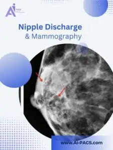

What Is Nipple Discharge?
Fluid may come from one or both nipples, either on its own or after pressure. Possible causes include pregnancy or breastfeeding, changes in the milk ducts, a benign growth called a papilloma, infection or some medicines. Cancer is another possible cause, so an unexplained change should be checked rather than diagnosed from its color.
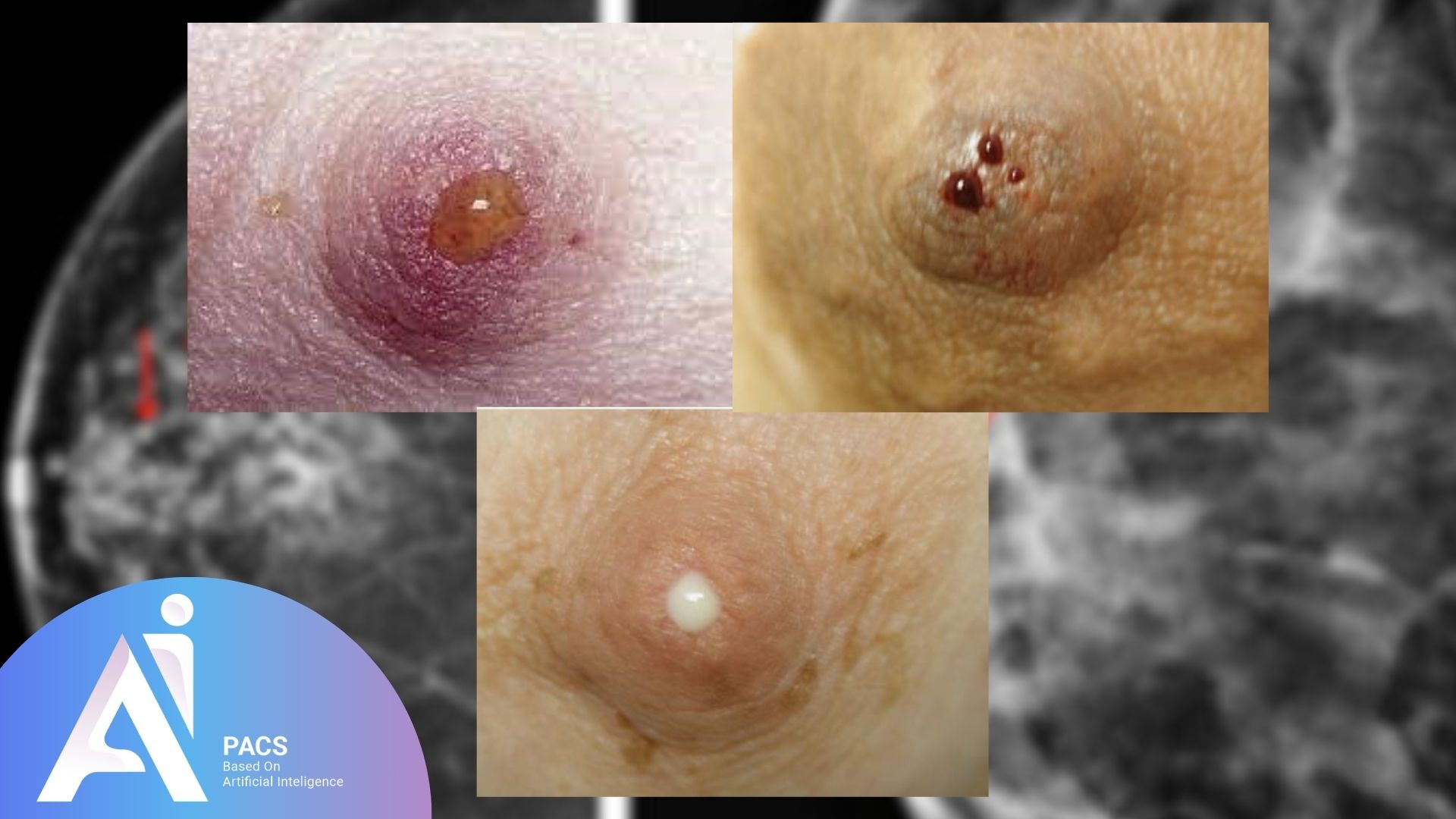
When Should You Seek Medical Advice?
Tell your clinician whether the discharge happens without squeezing, comes from one breast or one opening, and whether there is blood, a lump or a skin change. Discharge associated with squeezing and involving both breasts can have a different significance from spontaneous, one-sided discharge. Green fluid alone does not establish a dangerous cause, and clear fluid is not automatically reassuring. The complete pattern matters.
If discharge occurs during mammogram compression, tell the technologist, including whether it also happens at other times. Do not use that observation alone to decide whether evaluation is needed.
Which Imaging Test May Be Recommended?
For suspicious discharge, imaging is diagnostic rather than routine screening. In women under 30, ultrasound is usually the first test. At age 30 and above, diagnostic mammography or ultrasound may be appropriate, and the two may be used together. Your clinician chooses the investigation for your circumstances. Imaging is not routinely needed for a physiologic pattern when routine screening is current, but that distinction follows clinical assessment.
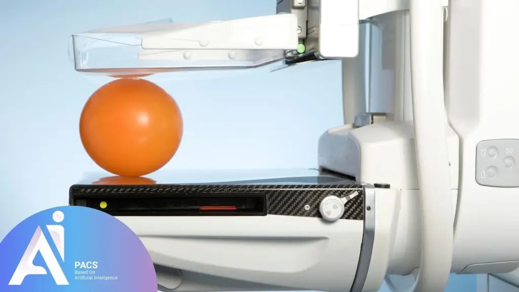
Mammography uses low-dose X-rays to show changes such as masses or calcifications. A normal mammogram does not necessarily explain the discharge or complete its evaluation. Depending on the findings, further assessment may include ultrasound, a biopsy or selected additional tests. A biopsy examines tissue; imaging alone does not provide every diagnosis.
Preparing for Diagnostic Mammography
Tell the imaging team about the discharge, any other breast changes, earlier procedures and possible pregnancy. Bring previous breast images and reports. Follow the center’s instructions about avoiding deodorant, powder or lotion on the breasts and underarms. A two-piece outfit can make changing easier. Tell the technologist if compression is painful. Do not postpone symptom assessment just to wait for a less tender point in your menstrual cycle.
Understanding the Result and Next Step
Read the report’s final assessment and recommendation together. Ask whether the imaging explains the symptom and what happens if it continues. The clinician may recommend follow-up, further imaging or tissue sampling. Treatment depends on the identified cause; a review of images does not prescribe treatment or replace a recommended biopsy.
When an Independent Image Review May Help
If you already have mammogram results and want the images reassessed, our mammogram second opinion page explains the review pathway. You can also start a consultation request and describe the question in your report.
Request the complete original DICOM study, relevant prior images and the written report from your imaging center. A report PDF or photograph alone is not enough for diagnostic image review. If your tests include ultrasound or another modality, ask the team to confirm the available files and review scope before ordering. A second read may agree with the original report.
Still waiting for a first report? Ask your imaging center when it will be available. For an alternative reporting request, read about online radiology reporting and check suitability. If you have symptoms but no images, arrange a clinical assessment first. Keep recommended appointments while arranging any independent review.