

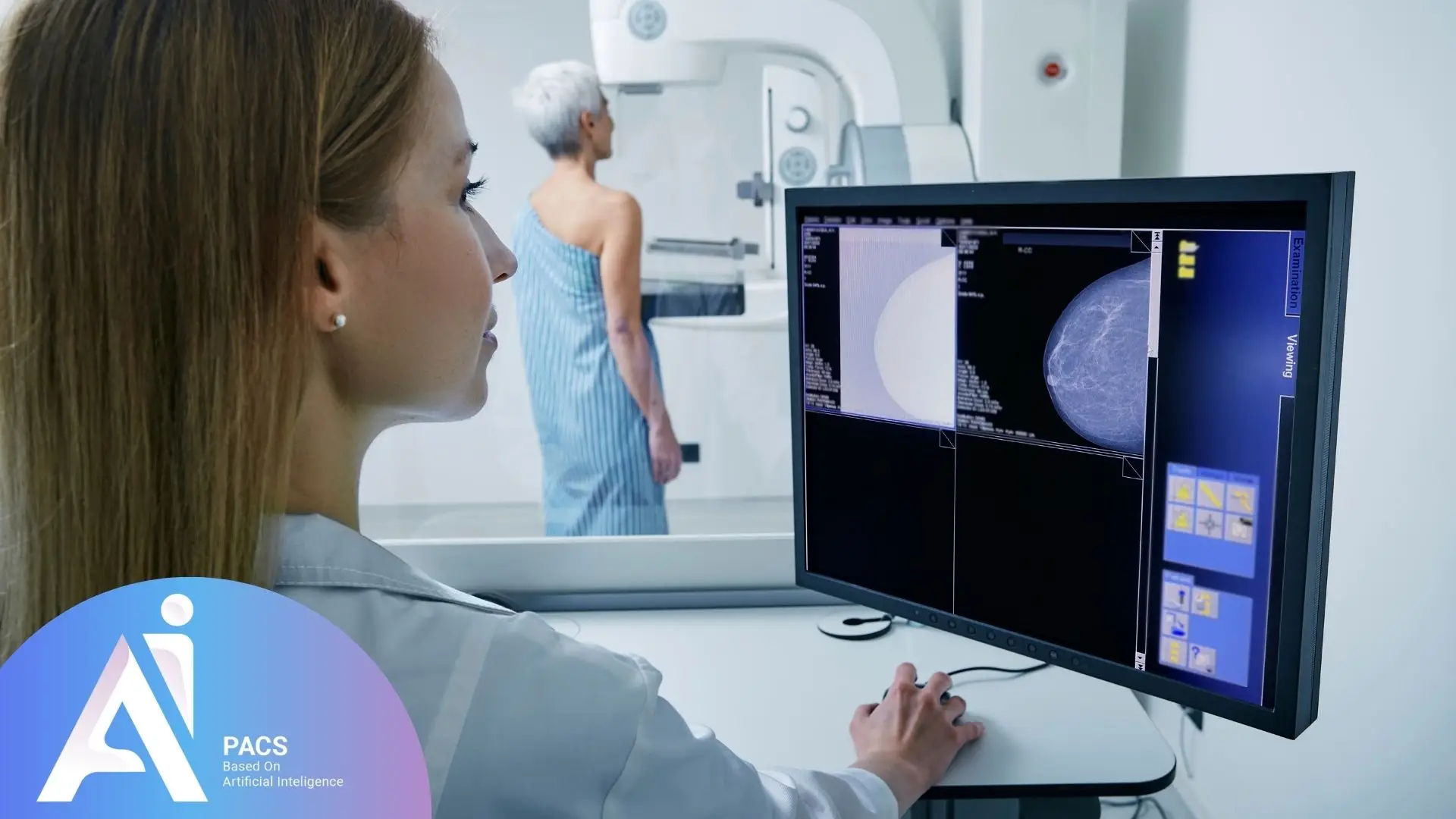
How to Read Your Mammogram Report
Start with the impression or conclusion, then read the recommendation. The finding, its assessment, and the next step belong together; one unfamiliar word does not explain the whole result.
- Check the exam details: Confirm the date and whether the study was screening or diagnostic.
- Look for comparison: Note whether previous mammograms were available.
- Read the findings: Identify the breast and location described, alongside the density statement.
- Find the final assessment: Read the BI-RADS category and the recommended action and timing.
For your appointment, write down: “My report describes ___; the assessment is ___; the recommended next step is ___.” If one of these is missing or unclear, ask your imaging team to explain it.
Want an independent image review? Learn about our mammogram second-opinion service or start a review request. Provide the original DICOM images, your report, and prior studies when available. Report text or screenshots alone are not enough for diagnostic image interpretation.
Specialized Terminology in Mammography
Understanding specialized terms in mammography is crucial for interpreting its results. For instance, knowing the meaning of ‘breast density’ can help patients understand why additional imaging might be required, while terms like ‘BI-RADS categories’ guide physicians in determining whether a biopsy is necessary, ultimately aiding in accurate diagnosis and treatment planning. Here are some common terms:
Terms Related to Images and Results
- Mammogram: An X-ray image of the breast taken during a mammography procedure.
- Breast Density: The amount of fibrous and glandular tissue compared with fat. Dense tissue can make findings harder to see, but density is not a cancer diagnosis.
- Mass: Any abnormal structure in the breast, which could be solid (like a tumor) or cystic (fluid-filled).
- Calcification: Small calcium deposits in breast tissue that appear as bright spots on a mammogram. These can be benign or indicative of cancer.
BI-RADS System
The Breast Imaging Reporting and Data System (BI-RADS) categorizes mammography findings to assess the risk of breast cancer. Categories range from 0 to 6:
- BI-RADS 0: Incomplete – additional evaluation needed.
- BI-RADS 1: Negative – no suspicious finding.
- BI-RADS 2: Benign findings.
- BI-RADS 3: Probably benign – requires short-term follow-up.
- BI-RADS 4: Suspicious abnormality – biopsy recommended.
- BI-RADS 5: Highly suspicious – biopsy strongly indicated.
- BI-RADS 6: Known malignancy already confirmed by biopsy.
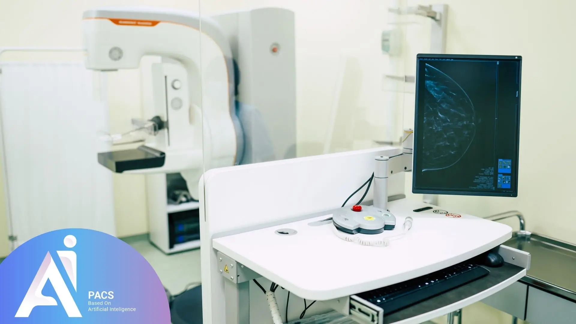
See our guide to BI-RADS categories for more detail. A category is an imaging assessment, not a cancer stage.
What Are Mammography Results?
Mammography results typically include radiographic images of breast tissue reviewed by a radiologist. After interpretation, the radiologist provides a detailed report that highlights any abnormalities, along with recommendations for follow-up actions or additional imaging if needed. This report is then shared with the patient’s physician, who discusses the findings and next steps with the patient.
Interpreting Mammography Results
Radiologists focus on specific indicators when interpreting mammography images:
- Masses and Nodules: These may be solid or cystic. Solid masses often require further examination.
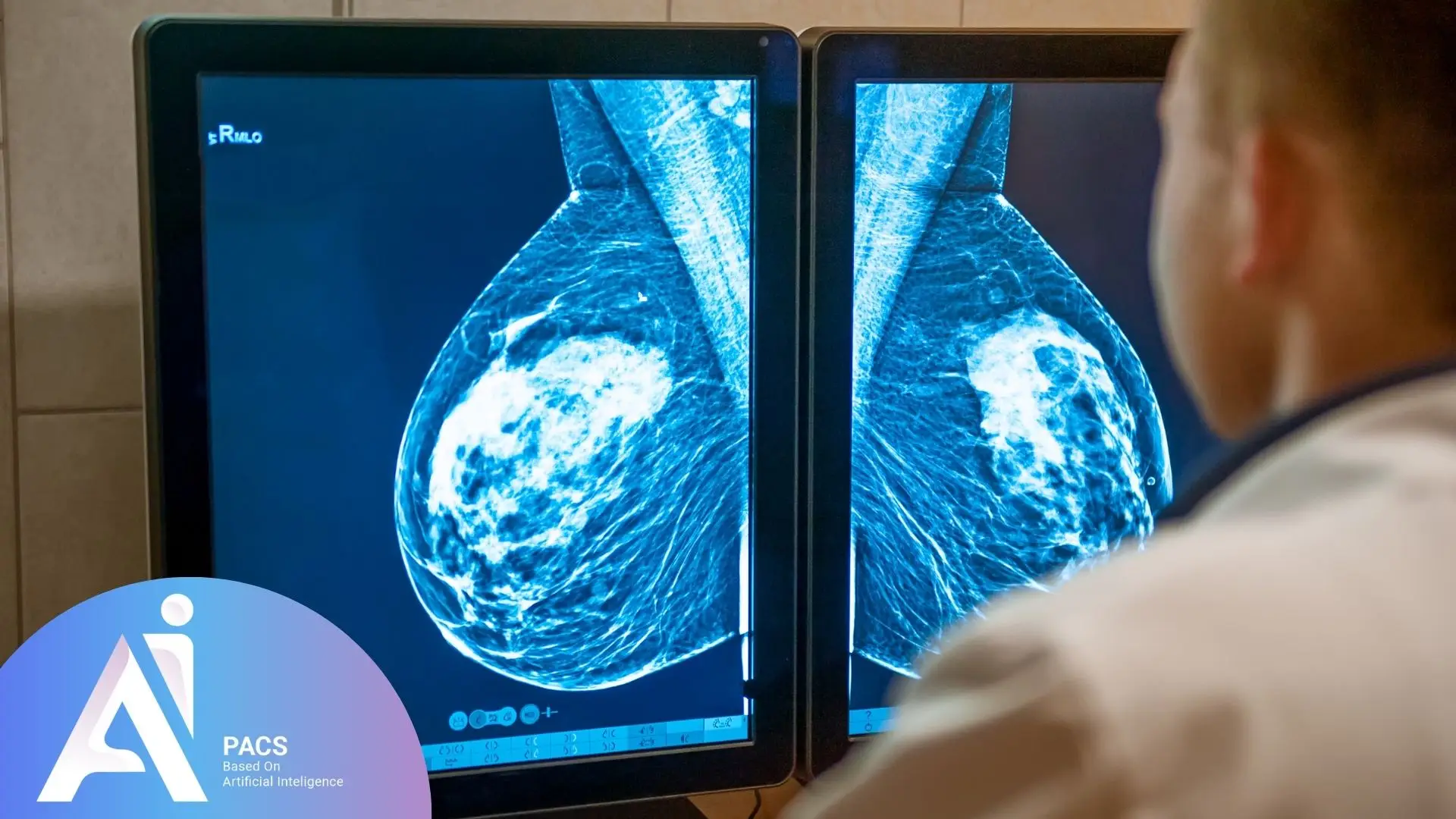
- Microcalcifications: These tiny calcium deposits are assessed by their shape, distribution, and any change over time. Some need additional imaging or biopsy; their presence alone does not diagnose cancer.
- Symmetry and Tissue Structure: Radiologists check for asymmetry or changes in the normal structure of breast tissue.
Follow-Up Is Different from Routine Screening
Follow the action and timing recommended for this examination. A callback may be needed for more images or comparison with older studies; it does not itself confirm cancer. Routine screening schedules depend on age, risk, and the guidance used by your care team. Do not substitute the next routine screening appointment for recommended diagnostic follow-up or biopsy.
Questions to Bring to Your Appointment
- Were my previous images available for comparison?
- Does the recommendation address a specific finding or my routine screening schedule?
- Who will arrange the next step, and when should I contact them if I have not heard back?
Have mammogram images but no report? Read about online radiology reporting or start a first-report request. The team needs the complete original DICOM study to assess the request.
Your Next Step
Keep a copy of your report and recommended follow-up plan. An independent image review can address questions about the imaging interpretation; it does not replace biopsy results or treatment advice from your treating team. Keep recommended appointments while arranging a review.
For a finding-specific explanation, read about mammography calcification and what it means.
References
- RSNA/ACR: How to read your mammography report
- RSNA/ACR: Dense breasts
- American Cancer Society: Understanding your mammogram report
- American Cancer Society: Mammograms
This educational guide does not replace medical assessment or the follow-up recommended by your care team.